Difficult Differentials in B-Cell Lymphomas Small B-Cell Lymphoma - - PowerPoint PPT Presentation

difficult differentials in b cell lymphomas small b cell
SMART_READER_LITE
LIVE PREVIEW

Difficult Differentials in B-Cell Lymphomas Small B-Cell Lymphoma - - PowerPoint PPT Presentation

5/24/2018 Difficult Differentials in B-Cell Lymphomas Small B-Cell Lymphoma with Increased Large Cells vs. Transformation to DLBCL Patrick Treseler, MD, PhD University of California San Francisco Small B-Cell Lymphoma with Increased Large


slide-1
SLIDE 1

5/24/2018 1

Patrick Treseler, MD, PhD University of California San Francisco

Difficult Differentials in B-Cell Lymphomas Small B-Cell Lymphoma with Increased Large Cells vs. Transformation to DLBCL

Small B-Cell Lymphoma with Increased Large Cells vs. DLBCL The Problem:

  • Small B-cell lymphomas (esp. MZL) can

have variable numbers of admixed large cells

  • How many large cells does it take to

indicate transformation to diffuse large B-cell lymphoma?

slide-2
SLIDE 2

5/24/2018 2

“MALT lymphoma, by definition, is a lymphoma composed predominantly by small cells. Transformed centroblast-like or immunoblast- like cells may be present in variable numbers, but when solid or sheet-like proliferations of transformed cells are present, the tumor should be diagnosed as diffuse large B-cell lymphoma (DLBCL) and the presence of accompanying MALT lymphoma noted.”

Transformation of Extranodal MZL (MALT lymphoma) to DLBCL

JR Cook et al. Extranodal marginal zone lymphoma of mucosa-associated lymphoid tissue (MALT lymphoma). In: WHO Classification of Tumours of Haematopoietic and Lymphoid Tissues (Swerdlow et al., eds). Lyon, France: International Agency for Research on Cancer, 2017, pp. 259-62.

Definition according to 2017 WHO…

slide-3
SLIDE 3

5/24/2018 3

CD21 CD21

slide-4
SLIDE 4

5/24/2018 4

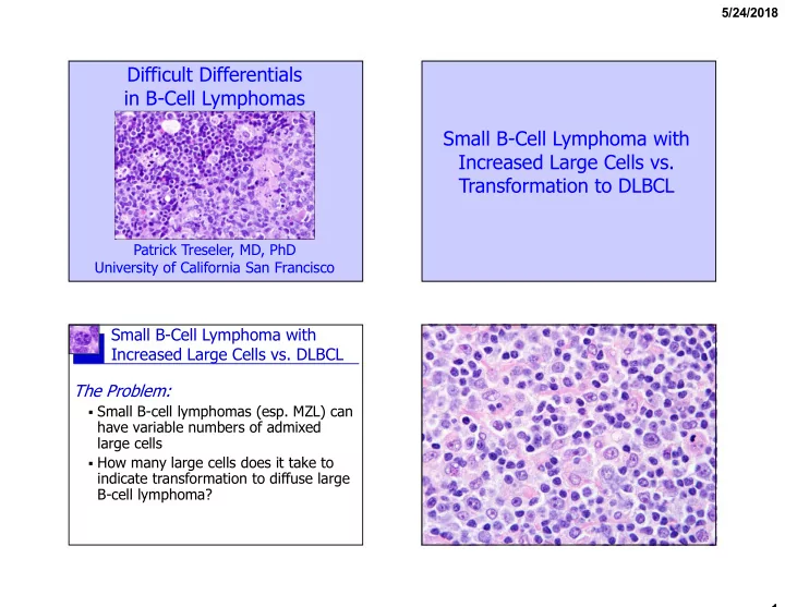
Small B-Cell Lymphoma with Increased Large Cells vs. DLBCL The Solution (for MALT lymphoma at least):

  • Solid clusters (dozens to hundreds?)
  • r diffuse sheets of large cells
  • Make sure you are not looking at a

B-cell follicle (use a CD21 stain)!

  • If large cells are increased but fall

short of clear-cut DLBCL, be descriptive & share your concern

“The presence of diffuse areas composed entirely or predominantly of large centroblasts (that would fulfil the criteria for grade 3 FL) in FL of any grade is equivalent to DLBCL, and a separate diagnosis of DLBCL should be made.”

Transformation of Follicular Lymphoma to DLBCL

ES Jaffe et al. Follicular lymphoma. In: WHO Classification of Tumours of Haematopoietic and Lymphoid Tissues (Swerdlow et al., eds). Lyon, France: International Agency for Research on Cancer, 2017, pp. 266-73.

Definition according to 2017 WHO…

  • Meets criteria for grade 3 FL
  • Architecture entirely diffuse (CD21)

Follicular Lymphoma

ES Jaffe et al. Follicular lymphoma. In: WHO Classification of Tumours of Haematopoietic and Lymphoid Tissues (Swerdlow et al., eds). Lyon, France: International Agency for Research on Cancer, 2017, pp. 266-73.

Grading per 2017 WHO

Grade* Definition

Grade 1-2 (low grade) 0-15 centroblasts/HPF 1 0-5 centroblasts/HPF 2 6-15 centroblasts/HPF Grade 3 (high grade) >15 centroblasts/HPF 3A Centrocytes present 3B Solid sheets of centroblasts

*WHO grading method derived from method of Mann and Berard (Mann RB, Berard CW. Criteria for the cytologic subclassification of follicular lymphomas: a proposed alternative method. Hematol Oncol. 1983 Apr-Jun;1(2):187-92.)

Grading of Follicular Lymphoma

  • Grade 1:

Practically all centrocytes; centroblasts present, but a little hard to find.

Cos Berard’s actual “eyeball” method (per Nancy Harris)

  • Grade 2:

Centroblasts easy to find, but mainly single scattered cells, and clearly in the minority

  • Grade 3:

Lots of centroblasts, often forming clusters

  • r sheets
slide-5
SLIDE 5

5/24/2018 5

FL, low grade (gr. 1-2) FL, grade 3A

slide-6
SLIDE 6

5/24/2018 6

CD21 CD21

FL, grade 3B

slide-7
SLIDE 7

5/24/2018 7

Small B-Cell Lymphomas

Basic Immunophenotypes

CD20 CD5 CD43 CD23 BCL1 BCL6 CD10

CLL/SLL + + + +

  • Mantle cell

+ + +

  • +
  • Follicular

+

  • /+
  • +

+/- Marginal +

  • /+
  • Proportion of cases positive: + >90%, +/- 50-90%, -/+ 10-50%, - <10%

t(11;14) Cyclin D1 “Histologically aggressive CLLs are recognized by proliferation centers that are broader than a 20x field or becoming confluent. Cases

Progression of CLL/SLL and its transformation to DLBCL

Campo et al. Chronic lymphocytic leukemia/small lymphocytic lymphoma. In: WHO Classification of Tumours of Haematopoietic and Lymphoid Tissues (Swerdlow et al., eds). Lyon, France: International Agency for Research on Cancer, 2017, pp. 216-21.

Definition according to 2017 WHO…

Full transformation to DLBCL is less precisely defined in the WHO, but would likely be best handled in a manner similar to that for extranodal marginal zone lymphoma.

Gine et al. Haematologica 95:1526; 2010

may also belong in this category when the Ki-67 index is >40% or there are >2.4 mitoses in the proliferation centers.”

Transformation of Mantle Cell Lymphoma to DLBCL

Swerdlow et al. Mantle cell lymphoma. In: WHO Classification of Tumours of Haematopoietic and Lymphoid Tissues (Swerdlow et al., eds). Lyon, France: International Agency for Research on Cancer, 2017, pp. 285-90.

Doesn’t happen!

But there are variants of mantle cell lymphoma that can be quite rich in large cells, and mimic DLBCL, specifically the pleomorphic variant. In the pleomorphic variant of MCL, the “cells are pleomorphic, but many are large,” and may have prominent nucleoli as well. Best gauge of prognosis in MCL is proliferative activity:

  • Worst prognosis: Ki-67 >30%, mitoses >50/mm2
  • Best prognosis: Ki-67 <10%, mitoses <25/mm2
slide-8
SLIDE 8

5/24/2018 8

CD20 CD5 BCL1

Mantle cell lymphoma, pleomorphic variant

slide-9
SLIDE 9

5/24/2018 9

Plasmacytoid Small B-Cell Lymphoma vs. Plasmacytoma

Plasmacytoid Small B-Cell Lymphoma vs. Plasmacytoma The Problem:

  • Plasmacytoid small B-cell lymphomas

(e.g., MZL) can have variable numbers

  • f mature plasma cells
  • If the proportion of plasma cells

approaches 100%, is it still a lymphoma, or is it a plasmacytoma?

slide-10
SLIDE 10

5/24/2018 10

CD20 Kappa

slide-11
SLIDE 11

5/24/2018 11

CD20 Kappa Lambda

Extramedullary plasmacytoma as form of marginal zone lymphoma? Hussong et al. (AJCP 111: 111; 1999)

  • Studied patients with extramedullary

plasmacytomas of GI tract, salivary gland, dura, and lymph nodes

  • All had lymphoepithelial lesions, if

epithelium present in tissues

  • All did well with resection ± radiation
  • Argued that extramedullary plasmacytoma

may be extreme form of MZL, distinct from plasmacytoma of bone and myeloma

Plasmacytoid Small B-Cell Lymphoma vs. Plasmacytoma The Solution:

  • Regard extraosseous tumors of mature

plasma cells as MZL/MALT lymphoma with extensive/extreme plasmacytic differentiation particularly if they:

  • Present in sites commonly involved by

MALT lymphoma

  • Have destructive lymphoepithelial lesions
  • But diagnose descriptively, encourage clinical

evaluation to exclude myeloma

slide-12
SLIDE 12

5/24/2018 12

Plasmacytoid Small B-Cell Lymphoma vs. Plasmacytoma

  • Plasmacytoid variants have been described for

essentially all small B-cell lymphomas

  • Look for expression of specific markers in the

residual small B-cells to rule those in:

  • CD5/CD23/LEF1  CLL/SLL
  • CD5/BCL1/SOX11  mantle cell lymphoma
  • CD10/BCL6  follicular lymphoma

And finally, keep in mind:

Plasmacytoma vs. Plasmablastic Lymphoma

Plasmacytoma vs. Plasmablastic Lymphoma

The Problem:

  • Plasmacytomas (e.g., in myeloma

patients) have variable numbers of plasmablasts (large neoplastic plasma cells with distinct to prominent nucleoli)

  • How many plasmablasts does it take to

call it plasmablastic lymphoma (DLBCL with plasma cell phenotype)?

slide-13
SLIDE 13

5/24/2018 13

Plasmacytoma vs. Plasmablastic Lymphoma (PBL)

The Solution: >50% (That was easy!)

  • Colomo et al. (AJSP 28:736) defined plasmablastic

lymphoma as subtype of DLBCL with:

  • Predominantly (>50%) large lymphoid cells (plasmablasts

and/or immunoblasts)

  • Plasma cell phenotype (CD20 neg/weakly pos., Oct-2 pos.)
  • Subtypes of PBL (per Colomo et al.)
  • PBL of oral mucosa type: All cells large
  • PBL with plasmacytic differentiation: Most cells large

immunoblasts or plasmablasts, but differentiation to small plasma cells present too

  • Extramedullary plasmablastic tumor: Identical to PBL with

plasmacytic diff, but history of prior or concurrent well differentiated plasma cell neoplasm (usually myeloma)

PBL, oral mucosa type PBL, oral mucosa type

slide-14
SLIDE 14

5/24/2018 14

PBL, with plasmacytic diff.

CD20 CD3 CD138

slide-15
SLIDE 15

5/24/2018 15

CD34 CD19 CD20 Pax-5 Surface Ig CD10 CD10 CD5 CD138 Oct-2

Germinal Center

Lymphoblastic leukemia/lymphoma CLL/SLL Mantle cell DLBCL? Follicular lymphoma Burkitt lymphoma DLBCL MZL DLBCL Plasmacytoma Plasmablastic lymphoma

Ig heavy chain genes rearranged Ig light chain genes rearranged Ig genes hypermutated

CD79a

Pax-5 CD20

slide-16
SLIDE 16

5/24/2018 16

Oct-2

Burkitt Lymphoma vs. Diffuse Large B-Cell Lymphoma

Burkitt Lymphoma vs. DLBCL

The Problem:

  • There are some B-cell lymphomas that

generally resemble BL, but either:

  • Have increased numbers of large

cells more typical of DLBCL; or

  • Lack the CD10+ BCL6+ BCL2-

phenotype of classical BL

  • What do we call these lymphomas?

BL? DLBCL? A BL/DLBCL gray zone? Something else?

Old WHO (2008):

Burkitt lymphoma B-cell lymphoma, unclassifiable, with features intermediate between DLBCL and Burkitt lymphoma (DLBCL/BL), a “gray zone” lymphoma (roughly corresponding to high-grade Burkitt-like lymphoma of 2001 WHO)

New WHO (2017):

Burkitt lymphoma DLBCL/BL “gray zone” cases all moved to new categories:

  • HGBL with rearrangements MYC and BCL2 +/- BCL6
  • HGBL, NOS
slide-17
SLIDE 17

5/24/2018 17

BL

Burkitt Lymphoma

  • Typical immunophenotype:

CD20 + CD10 + BCL6 + BCL2

  • CD10

BCL2 BCL6

*Weak BCL2+ seen in 20% Burkitt cases, does not exclude diagnosis of typical Burkitt lymphoma

Burkitt Lymphoma

  • Genotype:

Most have translocations linking MYC to Ig genes:

  • t(8;14) → MYC-IGH
  • t(2;8) → MYC-IGL-κ
  • t(8;22) → MYC-IGL-

*Up to 10% of otherwise typical Burkitt lymphoma may lack MYC translocations (11q aberration?)

Burkitt Lymphoma vs. “DLBCL/BL”

Acceptable for Classical Burkitt?

Morphology Immuno- phenotype

√ √ √ √

X X

Too many large cells

Not CD10+ BCL6+ BCL2-*

Burkitt Lymphoma “DLBCL/BL”

*Weak BCL2+ OK (seen in 20% Burkitt cases), & abnl IP can be redeemed by FISH MYC+ BCL2- BCL6-

slide-18
SLIDE 18

5/24/2018 18

MYC BCL2 BCL6

and/or

AJSP 29: 1652; 2005 P = 0.03 P = 0.003 P = 0.008 P = 0.32 (NS)

Snuderl et al. AJSP 34: 327; 2010

BL DLBCL Double Hit

Double hit lymphoma

  • DHL is a genotype associated with a very

poor prognosis in various B-cell neoplasms

  • Lymphomas with double-hit genotype:

– Burkitt or Burkitt-like lymphoma (most common) – Diffuse large B-cell lymphoma (less common) – Others (rare)

DLBCL/BL

slide-19
SLIDE 19

5/24/2018 19

DLBCL/BL

Burkitt Lymphoma vs. “DLBCL/BL”

Acceptable for Classical Burkitt?

Morphology Immuno- phenotype

√ √ √ √

X X

Too many large cells

Not CD10+ BCL6+ BCL2-*

Burkitt Lymphoma “DLBCL/BL”

*Weak BCL2+ OK (seen in 20% Burkitt cases), & abnl IP can be redeemed by FISH MYC+ BCL2- BCL6- Double hit lymphoma by FISH? HGBL with MYC and BCL2 and/or BCL6 rearrangements High-grade B-cell lymphoma, NOS

+

  • Swerdow et al. Blood 127:2375; 2016

Burkitt Lymphoma vs. DLBCL

The Solution:

  • If a B-cell lymphoma generally resembles

Burkitt lymphoma, but has either:

  • too many large cells for BL, or
  • an aberrant immunophenotype (not

CD10+ BCL-6+ BCL-2-), then:

  • Do FISH testing for translocations of

MYC, BCL2, and BCL6 genes, and place in appropriate subcategory of HGBL per 2017 WHO Classification

slide-20
SLIDE 20

5/24/2018 20