
SLIDE 1
1
Page 1 The Henry Moon Lecture The Continuing Dilemma of Ductal Carcinoma in Situ
Stuart J. Schnitt, M.D. Brigham and Women’s Hospital, Dana-Farber Cancer Institute, and Harvard Medical School Boston, MA
Disclosures
- None
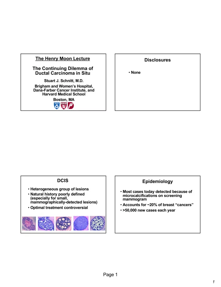
DCIS
- Heterogeneous group of lesions
- Natural history poorly defined
(especially for small, mammographically-detected lesions)
- Optimal treatment controversial
Epidemiology
- Most cases today detected because of
microcalcifications on screening mammogram
- Accounts for ~20% of breast “cancers”
- >50,000 new cases each year