computerized tomographic scan confirmed bilateral with
play

computerized tomographic scan confirmed bilateral with - PDF document

RESEARCH LETTER D ERMATOMYOSITIS AS THE I NITIAL P RESENTATION OF O VARIAN C ANCER Lai-Wan Chao, Lin-Hung Wei 1 * Departments of Obstetrics and Gynecology, and 1 Oncology, National Taiwan University Hospital and National Taiwan University


  1. ■ RESEARCH LETTER ■ D ERMATOMYOSITIS AS THE I NITIAL P RESENTATION OF O VARIAN C ANCER Lai-Wan Chao, Lin-Hung Wei 1 * Departments of Obstetrics and Gynecology, and 1 Oncology, National Taiwan University Hospital and National Taiwan University College of Medicine, Taipei, Taiwan. Dermatomyositis is a rare systemic inflammatory my- opathy with characteristic cutaneous manifestations consisting of heliotrope erythema, Gottron’s papules, poikiloderma, and periungual telangiectasia [1]. Diag- nosis is based on characteristic skin lesions and ad- ditional laboratory examination results. Laboratory confirmation of dermatomyositis includes increased lev- els of serum muscle enzymes (creatine phosphokinase, aldolase, and lactate dehydrogenase) and electromyo- graphic changes. Although the underlying pathogene- sis remains elusive, the association of this disease with malignancies is well known. Documented data from Scandinavia shows increased rates of malignant diseases in patients with dermatomyositis, compared with the Figure. Cutaneous lesions with generalized confluent macules general population [2,3]. We report here a case of epi- and violaceous erythema on the patient’s back. thelial ovarian cancer with an initial presentation of dermatomyositis. The manifestations of dermatomyosi- distension was present with no palpable abdominal tis regressed in synchrony with effective treatment of mass. On neurologic examination, the muscle power of the tumor with surgery and systemic chemotherapy. the bilateral arm, forearm and thigh muscles had de- A 60-year-old, multipara, menopausal woman, with creased to grade 4, while that of the unaffected muscles no remarkable medical history, presented to our derma- remained at grade 5. The results of laboratory examina- tology clinic with a complaint of intermittent facial tion revealed elevated levels of creatinine phosphokinase erythema for about 2 months. It was mainly distrib- (102 IU/L). The autoimmune disease profile was nor- uted on the bilateral cheeks and forehead. Progressive mal, except for antinuclear antibodies, which were pres- symmetrical muscle weakness, which led to difficulty ent at 1:160 + , in a speckle pattern. Myopathy was in cooking and climbing upstairs, was also reported, implied by electromyography results, which showed nu- together with progressive abdominal distension and in- merous small-amplitude and short-duration polyphasic creased abdominal circumference noted 10 days prior to waves with early recruitment in the right biceps. The skin admission. On physical examination, cutaneous lesions lesions and muscle weakness were only partially improved 
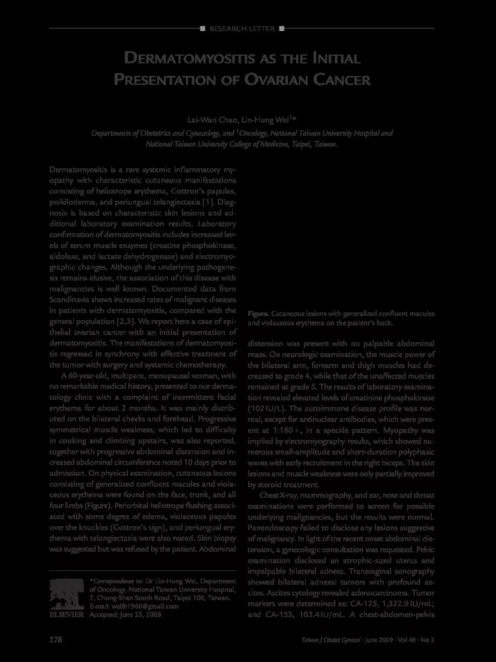
consisting of generalized confluent macules and viola- by steroid treatment. ceous erythema were found on the face, trunk, and all Chest X-ray, mammography, and ear, nose and throat four limbs (Figure). Periorbital heliotrope flushing associ- examinations were performed to screen for possible ated with some degree of edema, violaceous papules underlying malignancies, but the results were normal. over the knuckles (Gottron’s sign), and periungual ery- Panendoscopy failed to disclose any lesions suggestive thema with telangiectasia were also noted. Skin biopsy of malignancy. In light of the recent onset abdominal dis- was suggested but was refused by the patient. Abdominal tension, a gynecologic consultation was requested. Pelvic examination disclosed an atrophic-sized uterus and impalpable bilateral adnexa. Transvaginal sonography * Correspondence to: Dr Lin-Hung Wei, Department showed bilateral adnexal tumors with profound as- of Oncology, National Taiwan University Hospital, cites. Ascites cytology revealed adenocarcinoma. Tumor 7, Chung-Shan South Road, Taipei 100, Taiwan. markers were determined as: CA-125, 1,322.9 IU/mL; E-mail: weilh1966@gmail.com and CA-153, 103.4 IU/mL. A chest-abdomen-pelvis Accepted: June 25, 2008 178 Taiwan J Obstet Gynecol • June 2009 • Vol 48 • No 2

  2. Dermatomyositis and Ovarian Cancer computerized tomographic scan confirmed bilateral with dermatomyositis, there was a high incidence of ovarian solid tumors measuring 4.2 × 3.4 cm and ovarian cancer among the associated internal malig- 3.1 × 2.3 cm. Dirty infiltrations with increased soft tis- nancies (6/28) [5]. Nevertheless, the early detection of sue density on the mesentery and massive ascites were ovarian cancer among these patients is not substan- also noted. The patient was transferred to the gynecolo- tially higher. In a case series reported by Davis and gic ward owing to suggested ovarian malignancy. Ahmed [6], six dermatomyositis patients had abdomi- The patient underwent uncomplicated exploratory nal symptoms at presentation. All had undergone pre- laparotomy in which bilateral ovarian tumors with papill- vious screening for ovarian cancer, but ovarian disease ary growth and diffuse peritoneal seeding were observed. was not found in any of them. In the case series A total abdominal hysterectomy, bilateral salpingo- reported by Mordel et al [7], the diagnosis of derma- oophorectomy, infracolic omentectomy, and bilateral tomyositis preceded that of ovarian cancer in most cases, pelvic lymph node dissection were performed. Suboptimal with a mean interval of 10.9 months. Delayed diagnosis debulking surgery was achieved, and the residual tumor could be due to the insidious onset and slow progression measuring > 2 cm was mainly located in the perirectal of dermatomyositis and the limitations of imaging stud- area and over the visceral peritoneal surfaces. Results ies. Ninety-four percent of patients were diagnosed as of the final histopathologic examination confirmed a stage III or IV, rendering their prognosis extremely poor high-grade serous adenocarcinoma of the ovaries with [7]. Death from ovarian malignancies associated with omental metastasis, classified as International Federation dermatomyositis was 100%, and the mean survival time of Gynecology and Obstetrics stage IIIC ovarian can- from diagnosis was 11 months (range, 0–28 months) in cer. She completed five courses of adjuvant chemother- the series described by Davis and Ahmed [6]. Gynecolo- apy with paclitaxel (175 mg/m 2 ) and carboplatin (area gists should, therefore, be aware of the significance of under the curve, 6 mg/mL · min). The sixth course of the association between these two conditions. chemotherapy was not delivered because of impaired Some previous studies have described cases of der- liver function. The skin lesions and muscle weakness matomyositis presented after an established diagnosis resolved completely with the normalization of CA-125 of ovarian cancer [8]. However, as in the present case, levels. However, CA-125 rose again 5 months after the the diagnosis of ovarian cancer may occur shortly after initial chemotherapy, and the dermatomyositis reap- the diagnosis of dermatomyositis [9]. The risk of can- peared. Abdominopelvic magnetic resonance imaging cer was highest during the first year following the diag- revealed multiple enlarged lymph nodes in the bilateral nosis of dermatomyositis, and dropped substantially iliac chains. Salvage chemotherapy with liposomal thereafter. However, the risk of ovarian cancer did not doxorubicin (40mg/m 2 ) and carboplatin (area under the return to the expected population value for up to 5 years curve, 6 mg/mL · min) were prescribed for platinum- after diagnosis of dermatomyositis [1]. A thorough phy- resistant recurrent ovarian cancer. The symptoms of sical examination, pelvic ultrasound and serum CA-125 dermatomyositis resolved dramatically after the third assay should, therefore, be performed at the time of pre- course of salvage chemotherapy, and the patient was sentation and patients should be closely followed for still receiving treatment at the time of writing this report. several years. Dermatomyositis is a rare disease with an incidence In our patient, the manifestations of dermatomyosi- of 0.7–1.0/100,000 in the general population [4]. Its tis regressed in synchrony with effective treatment of the clinical significance lies in the fact that it is probably tumor. Although dermatomyositis may follow a para- a paraneoplastic event in some patients. A large retro- neoplastic course or may follow its own course, inde- spective study showed that up to 15% of dermatomyosi- pendent of tumor therapy [10], clinicians should be tis patients had underlying malignancies [3]. Ovarian, vigilant for the recurrence of muscle weakness and cuta- lung and colorectal cancers were frequently diagnosed neous manifestations that are associated with relapse both before and after the diagnosis of dermatomyositis, of the malignant disease in most cases. suggesting that these could be candidate cancers asso- In summary, the link between dermatomyositis and ciated with the disease. ovarian cancer should be clearly emphasized. As ovarian In a review of the literature, Cherin et al [5] reported cancer is the most lethal gynecologic cancer, clinicians six cases of dermatomyositis with ovarian cancer in a should perform timely screening in patients with der- series of 56 dermatomyositis patients (including 45 matomyositis to detect those with occult cancer, espe- women). The incidence of ovarian cancer in the female cially in female patients older than 40 years. Even if patients with dermatomyositis (13.3%) was much mortality cannot be prevented in these ovarian cancer higher than that observed in the general female popu- patients, disability from myositis can be alleviated when lation (1%). Moreover, in women older than 40 years the cancers are well managed. 179 Taiwan J Obstet Gynecol • June 2009 • Vol 48 • No 2

Download Presentation
Download Policy: The content available on the website is offered to you 'AS IS' for your personal information and use only. It cannot be commercialized, licensed, or distributed on other websites without prior consent from the author. To download a presentation, simply click this link. If you encounter any difficulties during the download process, it's possible that the publisher has removed the file from their server.

Recommend


More recommend