
SLIDE 1
7/21/2016 1
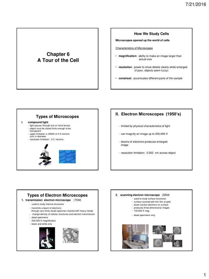
Chapter 6 A Tour of the Cell
How We Study Cells
Microscopes opened up the world of cells Characteristics of Microscopes
- magnification: ability to make an image larger than
actual size
- resolution: power to show details clearly while enlarged
(if poor, objects seem fuzzy)
- constrast: accentuates different parts of the sample
Types of Microscopes
I. compound light
- light passes through one or more lenses
- object must be sliced thinly enough to be
transparent
- upper limitation is 2000X or 0.5 microns
(um) in diameter
- resolution limitation: 0.2 microns
- II. Electron Microscopes (1950’s)
- limited by physical characteristics of light
- can magnify an image up to 200,000 X
- beams of electrons produces enlarged
image
- resolution limitation: 0.002 nm across object
Types of Electron Microscopes
- 1. transmission electron microscope (TEM)
- used to study internal structures
- transmits a beam of electrons
through very thinly sliced specimen stained with heavy metals
- change density of cellular structures and electron transmission
- dead specimens
- 200,000 X magnification
- black and white only
Plant Cell
2. scanning electron microscope (SEM)
- used to study surface structures
- surface covered with thin film of gold
- beam excites electrons on surface
- produces three dimensional images
- 100,000 X mag.
- dead specimens only