
SLIDE 1
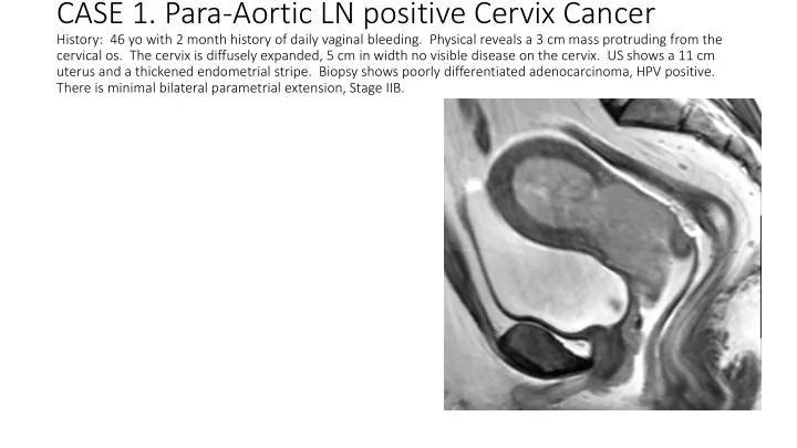
CASE 1. Para-Aortic LN positive Cervix Cancer
History: 46 yo with 2 month history of daily vaginal bleeding. Physical reveals a 3 cm mass protruding from the cervical os. The cervix is diffusely expanded, 5 cm in width no visible disease on the cervix. US shows a 11 cm uterus and a thickened endometrial stripe. Biopsy shows poorly differentiated adenocarcinoma, HPV positive. There is minimal bilateral parametrial extension, Stage IIB.