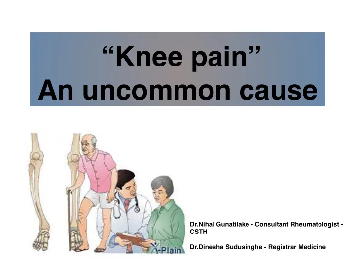

“Knee pain” An uncommon cause Dr.Nihal Gunatilake - Consultant Rheumatologist - CSTH Dr.Dinesha Sudusinghe - Registrar Medicine
Case history Mrs.J, 57 years P/C • B/L knee pain for 2 years H/P/C • Apparently healthy. • Progressive bilateral knee pain for the last 2 years. • Pain increases with activity and towards the end of the day. • Pain worst at night. • No associated swelling. • Neck pain and lower backache for the last 6 months. • No small joint pain or stiffness.
Case history … No heel pain, red eyes or scaly skin rash. • • Did not have alteration of bowel habits or lower urinary tract symptoms. LOA and LOW without evening pyrexia and drenching night sweats. • No pleuritic chest pain, chronic cough or contact history of TB. • Denied palpitation, easy fatiguability suggestive of anaemia. • Menopause 12 years back. • No family history of arthritis or malignancy. • PMHx - Not significant. Social Hx - Mother of three children. Activities of daily living — maintained (slowed).
?? Diagnosis • Middle aged female 1. KJ osteoarthritis • Non inflammatory KJ pain - Primary - Secondary • LOA and LOW 2. ?? Occult malignancy
Examination • Not pale. • No LN , clubbing • No goiter/breast lumps
Examination… Cardiovascular Respiratory - BP-130/90mmHg - No added sounds - PR- 88 bpm - Apex - normal position - No murmur
Examination… • Abdomen - No organomegaly • Neurology - Cranial nerves - normal - No motor weakness - No sensory impairment
Musculoskeletal examination B/L KJ • No deformity • No swelling Early osteoarthritis • Full range of movements • Crepitus + Spine • No deformities or muscle spasms • Full range of movement without pain
What is the diagnosis? 1. KJ osteoarthritis - Primary - Secondary 2. ?? Occult malignancy
X-ray knee joint - sclerotic lesions
X-ray cervical spine - lateral view Sclerosis of cervical vertebrae
Sclerotic bone lesions Focal or multifocal sclerotic bone lesions Diffuse Sclerotic Bone Lesions • Vascular •Vascular ◦ Hemangioma ◦ Infarct (e.g. sickle cell) ◦ Infarct •Neoplasm • Infection ◦ Metastatic ◦ Chronic osteomyelitis ▪ Prostate • Neoplasm ▪ Breast ◦ Primary ▪ Osteosarcoma •Drugs ◦ Metastatic ◦ Vitamin D • Trauma ◦ Fluoride ◦ fracture (stress) •Endocrine/Metabolic • Endocrine/Metabolic ◦ Hyperparathyroidism ◦ Paget's disease
D/D • Metastatic bone disease • Paget’s disease • Osteoblastic lymphoma • KJ osteoarthritis - Primary - Secondary
Investigations • FBC WBC 5.8 x Hb 12.8 g/dl Normal PLT 230 x 103 • ESR 15 mm 1st hr • S.Ionized Ca2+ 1.13 mmol/l • S.Phosphate 4 mg/dl
Investigations • LFT ALT 28 U/L AST 38 U/L ALB 58 mg/dl Total protein 70 mg/dl Isolated elevation of ALP ALP 1726 U/L GGT 38 U/L TBIL 14 µmol/l • Scr 60 µmol/l • USS abdomen • No organomegaly • No intra abdominal lymphadenopathy
Skull x-ray - lateral “Cotton wool” skull
X-ray pelvis - lytic and sclerotic lesions Cortical thickening
Diagnosis • Metastatic bone disease • Paget’s disease • Osteoblastic lymphoma • KJ osteoarthritis - Primary - Secondary
Paget’s disease • Sir James Paget first described chronic inflammation of bone as osteitis deformans in 1877. • Today it is known as, Paget’s disease of bone.
Paget's disease • Second most common bone disorder (after osteoporosis) in elderly. • Common among male. • Cause unknown. • Chronic, progressive disorder. • Localized area of excessive bone resorption and formation. • Frequently multifocal. • New lesions rarely develop in previously un affected areas after the diagnosis.
Paget's disease • Predilection for the axial skeleton. (pelvis, femur, lumbar spine, and skull) (descending order of frequency)
Pathophysiology Normal Paget’s
Pathophysiology Three phases 1. Lytic phase 2. Mixed phase 3. Sclerotic phase At any one time, multiple stages of the disease may be demonstrated in different skeletal regions at different rates of progression.
Histology
Clinical features • Majority are asymptomatic. • Patient may present with non-specific symptoms or symptoms suggestive of another disease, Bone pain Osteoarthritis Deformity Fracture Deafness • Diagnosis is often based on incidental findings Elevated total or bone specific ALP Radiological findings
Examination Facial disfiguration Skull enlargement Bowing of long bones
Diagnosis • Serological investigations - Total alkaline phosphatase (ALP) - Bone specific ALP • Radiograph - characteristic appearance • Bone scan - to assess the extent of the disease
Radiological investigations - Lytic phase Osteoporosis circumscripta ‘V’ shaped “blade of grass” lesion
Sclerotic phase
Advanced paget’s disease sclerotic and lytic lesions “Cotton wool” skull
Paget’s disease of vertebra - picture frame vertebral body Cortical thickening
Bone scan - polyostotic disease
Complications Acceletated bone remodeling Osteosarcoma Impaired bone Bone enlargement Hypervascularity micro architecture Bony overgrowth Micro-fractures Fractures High output cardiac around nerves failure Bowing deformity of weight bearing bones Nerve impingement syndrome Secondary Gait change and osteoarthritis mechanical stress Back pain and joint pain
Treatment Indications Metabolically active disease • Bone pain • Fracture • Bony deformities and weight-bearing bone involvement. • Compression of spinal cord or nerve roots
Treatment Preparation for orthopedic surgery. (If joint replacement anticipated at involved site within 6 months) Hypercalcaemia or hypercalciuria - recurrent renal calculi. Serum ALP levels greater than twice the upper limit of the reference range.
Treatment Non - pharmacological • Gait abnormality - canes and walkers Pharmacological • Bisphosphonate • NSAID/Opioid - pain management Surgery • Bone deformities, fractures or secondary osteoarthritis
Bisphosphonates Bisphosphonates • Antiresorptive agent - osteoclast apoptosis • Inhibit bone turnover • Improve bone pain • C/I if GFR < 35 ml/min - can substitute with calcitonin
Compare the effects of two management strategies on fracture, quality of life, bodily pain, and other common complications of PDB, including the requirement for orthopedic surgery and hearing loss. Symptomatic Intensive Any fracture 7.4% 7.0% Pagetic bone pain 30.8% 26.4% Any bone pain 73.7% 69.7% Quality of life -1.2% -1.3%
Bisphosphonates… Drug Dose Fall in ALP Reference 40mg/day, orally, 73-79% in 6 Alendronate Siris 1996 for 6 months months 30mg/day, orally, Risedronate 69% in 6 months Reid 1996 for 2 months 60mg/day, Pamidronate 53% in 6 months Miller 2004 intravenously, for 3days 5mg, Zolendronic 80% in 6 months Reid 2005 intravenously, single dose
Follow up • Serum total or bone specific ALP - fall within 7-10 days of starting treatment and nadir after 3-4 months. • ALP every 1-2 years in zolendronic acid treated group • Periodic x-rays of osteolytic lesions. Retreatment indicated if patient has not responded after 6 months of treatment or clinical or biochemical relapse.
Surgical treatment Corrective osteotomy for deformity Hip replacement
Our patient.. • Alandronate 70 mg EOD • Awaiting Zolendronic acid
Future … . ZiPP (Zoledronate in Prevention of Paget’s disease) Randomized trial of genetic testing and targeted zolendronic acid therapy to prevent SQSTM1 mediated Paget’s disease.
Take home message • Morbidity from Paget’s disease can be extensive. • Most of the patients are asymptomatic at presentation. • Important to suspect and initiate treatment early to prevent complications. • Treatment does not cure the disease, but it can control. • Prognosis is good, if treatment administered before major changes have occurred.
References • Singer FR, Bone HG, Hosking DJ, Lyles KW, Murad MH, Reid IR, Siris ES, Endocrine Society. Paget's disease of bone: an endocrine society clinical practice guideline . J Clin Endocrinol Metab. 2014 Dec;99(12):4408-22. • Guideline Stresses Bisphosphonate Infusion for Paget ...www.medscape.org/viewarticle/837040 • Ralston SH, Layfield R. Pathogenesis of Paget disease of bone . Calcif Tissue Int 2012;91:97-113. • Stuart H. Ralston, M.D. Paget's disease of bone . N Engl J Med 2013; 368:644-650. • Siris ES, Roodman GD. Paget's disease of bone . In: Rosen C, ed. Primer on the metabolic bone diseases and disorders of mineral metabolism. Hoboken, NJ: Wiley, 2012:335-43. • Reid IR, Lyles K, Su G, et al. A single infusion of zoledronic acid produces sustained remissions in Paget disease -- data to 6.5 years. J Bone Miner Res 2011;26:2261-2270 • Langston AL, Campbell MK, Fraser WD, MacLennan GS, Selby PL, Ralston SH. Randomized trial of intensive bisphosphonate treatment versus symptomatic management in Paget's disease of bone. J Bone Miner Res 2010;25:20-31
Recommend
More recommend