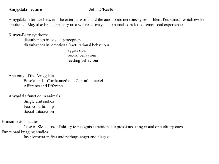
SLIDE 1 Amygdala lecture John O’Keefe Amygdala interface between the external world and the autonomic nervous system. Identifies stimuli which evoke
- emotions. May also be the primary area where activity is the neural correlate of emotional experience.
Kluver-Bucy syndrome disturbances in visual perception disturbances in emotional/motivational behaviour aggression sexual behaviour feeding behaviour Anatomy of the Amygdala Basolateral Corticomedial Central nuclei Afferents and Efferents Amygdala function in animals Single unit sudies Fear conditioning Social Interaction Human lesion studies Case of SM - Loss of ability to recognise emotional expressions using visual or auditory cues Functional imaging studies Involvement in fear and perhaps anger and disgust
SLIDE 2
SLIDE 3 Kluver-Bucy Syndrome following bilateral temporal lobectomy in monkeys. Main components are: visual defects,
changes in emotional behaviour (hypersexuality,hypo-emotionality)
SLIDE 4
Basolateral Corticomedial Central
Primate amygdala Transverse sections
SLIDE 5 Central nucleus visceral visceral
Cortico- medial nuclei hypothalamus neocortex Basolateral nuclei dorsomedial nucleus of the thalamus Prefrontal cortex Input Amygdala nucleus Output
SLIDE 6 Amygdala inputs and
SLIDE 8
SLIDE 9
visual input
SLIDE 10
SLIDE 11
Amygdala efferent-Stria Terminalis
SLIDE 12
Amygala Eating Cells
Liu et al 2018
SLIDE 13
Liu et al 2018
Habituation in Eating Cells
SLIDE 14
SLIDE 15
Amygdala cells are Specific Responsive 55% One Stimulus 61% Rat 40% Food 35% Transport Box 18%
SLIDE 16
Other selective amygdala cells
SLIDE 17
selective rat cells
SLIDE 18
sec
active memory traces for eating rice
SLIDE 19 active memory traces for experience
transport box
SLIDE 20
active memory traces are independent of behavior
SLIDE 21
Pavlovian fear conditioning
SLIDE 22
Nuclei involved in conditioned fear and their autonomic and behavioural actions
SLIDE 23
SLIDE 24
Effect of amygdala lesions on social behavior
SLIDE 25
SLIDE 26
affiliative vocalizations towards mother/infant pairs are reduced by amygdala lesions
SLIDE 27
Ekman Emotional faces
SLIDE 28
Ekman Emotional faces
SLIDE 29
electrode placements in temporal lobe
SLIDE 30 human amygdala cell responding to emotion
SLIDE 31 Mormann 2011
right human amygdala cells responding to pictures
SLIDE 32
SM’s Missing amygdala
SLIDE 33 S.M’s drawings
emotions
SLIDE 34
SLIDE 35
SLIDE 36
Bilateral amygdala activation from threatening words