SLIDE 1
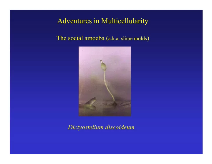
Adventures in Multicellularity
The social amoeba (a.k.a. slime molds) Dictyostelium discoideum
SLIDE 2 Dictyostelium discoideum
- the most studied of the social amoebae / cellular slime molds
- predatory soil amoeba that feeds on a variety of microorganisms and
decaying matter
(in the lab exist primarily on a diet of E. coli although strains have been selected that grow on complex media alone).
- asexually reproducing amoebae (unicellular) under conditions when food is
abundant
- under starvation conditions a complex developmental cycle is initiated – an
adventure in multicellularity.
- they have features of both plants and animals
- cellulose and development of spores (plant-like)
- cell movements involved in morphogenesis (animal-like)
SLIDE 3 Embryogenesis Dictyostelium Development
Aggregation of many identical cells Successive divisions
cell (zygote)
SLIDE 4
The Natural History of Dictyostelium discoideum
Vegetative Cycle: Amoeba lives solitary existence feeding on bacteria Reproduce by binary fission (i.e. divide in half) Continues as long as food is available
Feeding is by phagocytosis – essentially engulfing their prey with membrane. RH Kessin (2001)
SLIDE 5
The Natural History of Dictyostelium discoideum
Chemotaxis and Aggregation: When the cells begin to starve, they begin a developmental cycle They produce and release cAMP that acts as a chemoattractant and also as a signaling molecule The cells comes together (streaming) to form a multicellular tipped aggregate . This elaborates into a finger or standing slug
RH Kessin (2001)
SLIDE 6
The Natural History of Dictyostelium discoideum
The standing slug has two fates – it can topple over and become a migrating slug or it can proceed to develop into a fruiting body. The elaboration of the fruiting body begins with culmination
RH Kessin (2001)
SLIDE 7
The Natural History of Dictyostelium discoideum
Scanning electron micrographs of the different stages of development
RH Kessin (2001)
SLIDE 8 The Natural History of Dictyostelium discoideum
1. 2. 3. 4.
- 1. Chemotaxis to signals produced
coordinately by the cells (primitive morphogens)
- 2. Aggregation and cell adhesion
(primitive patterning and multicellularity)
- 3. Differentiation and patterning in
the migrating slug and fruiting body (primitive polarity)
- 4. Competition to be spores
(cheating)
RH Kessin (2001)
SLIDE 9
The Natural History of Dictyostelium discoideum
Enclosed in a sheath (muco- polysaccharide and cellulose) The tip is a specialized group of cells that control development
(remove it and development stops until a new tip is formed)
Under the control of the tip the aggregate elongates and forms the standing slug.
RH Kessin (2001)
SLIDE 10
The Natural History of Dictyostelium discoideum
The slug typically contains about 100,000 cells and behaves as a single entity. It is capable of both phototaxis and thermotaxis.
RH Kessin (2001)
SLIDE 11 Dictyostelium aggregates have many properties of an embryo :
- They are essentially a multicellular entity enclosed within a sheath
- They have polarity
- They have exquisite proportioning (1/5 prespore ; 4/5 prestalk)
- They have an organizing center (tip)
- morphogenesis involves cell movements
SLIDE 12
The essential steps in cell motility
RH Kessin (2001)
SLIDE 13
Three dimensional reconstruction of a D. discoideum amoebae moving towards a cAMP gradient.
cAMP source cAMP
Wessel and Soll, Univ. of Iowa
SLIDE 14 Chemotaxis of single cell towards a point source of cAMP.
The tip of the capillary tube is the source of cAMP. See how quickly the cell responds to changes in concentration. Note the number of pseudopodia protrusions in the amoeba.
Time hr:min:sec:
- G. Gerisch, Max Planck Institute, GDR
SLIDE 15
- D. discoideum amoebae chemotaxing toward cAMP
Cells are expressing a GFP-coronin fusion protein: coronin is a cytoskeletal protein that is localized in the extended pseudopods.
- G. Gerisch, Max Planck Institute, GDR
SLIDE 16
Starvation and cAMP signaling
During starvation, cells develop the ability to synthesize, detect and degrade cAMP. When one cell releases a pulse of cAMP (1), neighboring cells detect it and move up the gradient towards the source (2) . After a delay of about one minute these cells release cAMP of there own (2) and process gets repeated about every six minutes (3). This process results in waves of migration (4). 1 2 3 4
RH Kessin (2001)
SLIDE 17
Aggregation patterns
The patterns of aggregation can be seen by dark-field microscopy because moving and stationary cells reflect light differently – moving cells appear lighter.
Note the waves of cAMP tend to propagate from a central point (‘organizing center’) RH Kessin (2001)
SLIDE 18
Dark waves of D. discoideum cells on caffeine agar. Movie constructed from 36 seconds intervals. (F. Siegert and C.J. Weijer J.Cell Sci. 93:325-335 (1989))
Aggregation Patterns
SLIDE 19
Core of a Spiral Wave in Aggregating D. discoideum cells
10 sec between images
Siegert and Weijer, J. Cell Sci. 93:325-335.
SLIDE 20
Siegert and Weijer, Curr Biol 1995
Rotation of cells within the mound.
SLIDE 21
The classical studies of Raper (1940’s)
RH Kessin (2001)
Prestalk and prespore cells do not mix throughout development.
SLIDE 22
The classical studies of Raper (1940’s)
RH Kessin (2001)
The tip controls events in the slug.
Apical fractions from foreign slugs (B-D) were grafted onto a normal slug. Four independent slugs emerge after ~45 min.
SLIDE 23
JT Bonner (2000)
Positional Sorting of Prestalk and Prespore cells within the slug
Prestalk cells were grafted onto the anterior end of a slug. These cells eventually migrate to the proper position at the posterior end of the slug.
SLIDE 24
JT Bonner (2000)
There is little mixing of prestalk and prespore cells in the slug.
The prestalk cells at the tip move more rapidly in the slug than the pre-spore cells.
Prestalk Prespore This study used an ingenious method developed by Bonner to generate “two dimensional” slugs. They could contain as few as 50 cells (typical slugs have > 100,000 cells) but the correct proportioning of prestalk and prespore cells was always maintained.
SLIDE 25
Wolpert, 1998
Movement of Cell Types During Culmination
SLIDE 26
Weijer, Univ. of Dundee
Note that cells move more rapidly in the tip (prestalk cells) than in the body of the slug (prespore cells). Note also that the tip is further partitioned into Prestalk a nd Prestalk O cells.
Movement of Prestalk Cells in the Slug
Two types of prestalk cells: 1) Green (GFP labeled) Prestalk A cells 2) Red (red-shifted GFP labeled) Prestalk O cells
SLIDE 27
Weijer, Univ. of Dundee
Movement of Prestalk Cells During Culmination
Two types of prestalk cells: 1) Green (GFP labeled) Prestalk A cells 2) Red (red-shifted GFP labeled) Prestalk O cells Watch closely for the movement of prestalk cells down the core of the culminating slug.
SLIDE 28 Mound formation can be described by two assumptions: 1.) by considering the population of cells as an excitable medium and 2.) by considering cell movement as the flow of a compressible liquid. The cell movement (fluid flow) is controlled by chemotactic forces dependent on increasing concentrations of the cAMP. The model allows to describe the whole early development from isolated single cells into bifurcating aggregation streams to the formation of a 3-dimensional, hemispherical mound, (see Vasiev B., Siegert F. & C. J.Weijer (1997), J.Theor.
SLIDE 29 A three dimensional hybrid cellular automata/partial differential equation model capable of describing the morphogenesis of Dictyostelium discoideum “from single cells to crawling slug". Using simple local interactions we can achieve the morphogenesis with only three processes: production of and chemotaxis to cAMP and cellular adhesion. The interplay of these processes causes the amoebae to spatially self-organize leading to the complex behaviour
- f stream and mound formation, cell sorting and slug migration all
without any change of parameters. Our model describes the substrate for morphogenesis on which higher cell functions can
Savill and Hogeweg (1997) J. Theor. Biol. 184:0229-235. http://www-binf.bio.uu.nl/~njs/dicy.html
- P. Hogeweg et al Model of Development (Univ. of Utrecht)
SLIDE 30 Streaming and Mound Formation
Hogeweg Model (cont’d)
- Each amoeba is represented by connected automata in CA system (not
just as a single cell)
- Cell-cell adhesion an chemotaxis are represented as simple CA rules
- Amoeba are not rigidly defined by the number of cells but can fluctuate
slightly allowing flexible interactions and movements past one another
SLIDE 31
cAMP waves Slug development
Hogeweg Model (cont’d)
Yellow Prestalk Green Prespore
SLIDE 32
Siegert and Weijer, J. Cell Sci. 93:325-335.
SLIDE 33
General References:
First Signals : Evolution of Multicellular Development. J.T. Bonner (2000) Princeton University Press Dictyostelium: Evolution, Cell Biology and the Development of Multicellularity. Richard H. Kessin (2001) Cambridge University Press Web sites containing numerous links to research sites, resources and databases: http://dicty.cmb.nmu.edu/dicty/dicty.html http://dictybase.org/dicty.html