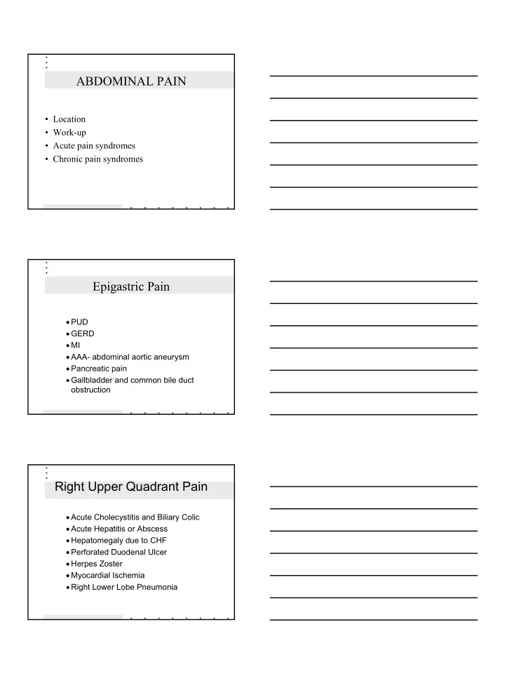
abdominal pain
play

ABDOMINAL PAIN Location Work-up Acute pain syndromes Chronic - PDF document

ABDOMINAL PAIN Location Work-up Acute pain syndromes Chronic pain syndromes Epigastric Pain PUD GERD MI AAA- abdominal aortic aneurysm Pancreatic pain Gallbladder and common bile duct obstruction Right


  1. ABDOMINAL PAIN • Location • Work-up • Acute pain syndromes • Chronic pain syndromes Epigastric Pain • PUD • GERD • MI • AAA- abdominal aortic aneurysm • Pancreatic pain • Gallbladder and common bile duct obstruction Right Upper Quadrant Pain • Acute Cholecystitis and Biliary Colic • Acute Hepatitis or Abscess • Hepatomegaly due to CHF • Perforated Duodenal Ulcer • Herpes Zoster • Myocardial Ischemia • Right Lower Lobe Pneumonia 1

  2. Left Upper Quadrant Pain • Acute Pancreatitis • Gastric ulcer • Gastritis • Splenic enlargement, rupture or infarction • Myocardial ischemia • Left lower lobe pneumonia Right lower Quadrant Pain • Appendicitis • Regional Enteritis • Small bowel obstruction • Leaking Aneurysm • Ruptured Ectopic Pregnancy • PID • Twisted Ovarian Cyst • Ureteral Calculi • Hernia Left Lower Quadrant Pain • Diverticulitis • Leaking Aneurysm • Ruptured Ectopic pregnancy • PID • Twisted Ovarian Cyst • Ureteral Calculi • Hernia • Regional Enteritis 2

  3. Periumbilical Pain • Disease of transverse colon • Gastroenteritis • Small bowel pain • Appendicitis • Early bowel obstruction Diffuse Pain • Generalized peritonitis • Acute Pancreatitis • Sickle Cell Crisis • Mesenteric Thrombosis • Gastroenteritis • Metabolic disturbances • Dissecting or Rupturing Aneurysm • Intestinal Obstruction • Psychogenic illness Referred Pain • Pneumonia (lower lobes) • Inferior myocardial infarction • Pulmonary infarction 3

  4. TYPES OF ABDOMINAL PAIN • Visceral – originates in abdominal organs covered by peritoneum • Colic – crampy pain • Parietal – from irritation of parietal peritoneum • Referred – produced by pathology in one location felt at another location ORGANIC VERSUS FUNCTIONAL PAIN HISTORY ORGANIC FUNCTIONAL Pain character Acute, persistent pain Less likely to change increasing in intensity Pain localization Sharply localized Various locations Pain in relation to sleep Awakens at night No affect Pain in relation to Further away At umbilicus umbilicus Associated symptoms Fever, anorexia, Headache, dizziness, vomiting, wt loss, multiple system com- anemia, elevated ESR plaints Psychological stress None reported Present WORK-UP OF ABDOMINAL PAIN HISTORY • Onset • Qualitative description • Intensity • Frequency • Location - Does it go anywhere (referred)? • Duration • Aggravating and relieving factors 4

  5. WORK-UP PHYSICAL EXAMINATION • Inspection • Auscultation • Percussion • Palpation • Guarding - rebound tenderness • Rectal exam • Pelvic exam WORK-UP LABORATORY TESTS • U/A • CBC • Additional depending on rule outs – amylase, lipase, LFT’s WORK-UP DIAGNOSTIC STUDIES • Plain X-rays (flat plate) • Contrast studies - barium (upper and lower GI series) • Ultrasound • CT scanning • Endoscopy • Sigmoidoscopy, colonoscopy 5

  6. Common Acute Pain Syndromes • Appendicitis • Acute diverticulitis • Cholecystitis • Pancreatitis • Perforation of an ulcer • Intestinal obstruction • Ruptured AAA • Pelvic disorders APPENDICITIS • Inflammatory disease of wall of appendix • Diagnosis based on history and physical • Classic sequence of symptoms – abdominal pain (begins epigastrium or periumbilical area, anorexia, nausea or vomiting – followed by pain over appendix and low grade fever DIAGNOSIS • Physical examination – low grade fever – McBurney’s point – rebound, guarding, +psoas sign • CBC, HCG – WBC range from 10,000-16,000 SURGERY 6

  7. DIVERTICULITIS • Results from stagnation of fecal material in single diverticulum leading to pressure necrosis of mucosa and inflammation • Clinical presentation – most pts have h/o diverticula – mild to moderate, colicky to steady, aching abdominal pain - usually LLQ – may have fever and leukocytosis PHYSICAL EXAMINATION • With obstruction bowel sounds hyperactive • Tenderness over affected section of bowel DIAGNOSIS • Often made on clinical grounds • CBC - will not always see leukocytosis MANAGEMENT • Spontaneous resolution common with low-grade fever, mild leukocytosis, and minimal abdominal pain • Treat at home with limited physical activity, reducing fluid intake, and oral antibiotics (bactrim DS bid or cipro 500mg bid & flagyl 500 mg tid for 7-14 days) • Treatment is usually stopped when asymptomatic • Patients who present acutely ill with possible signs of systemic peritonititis,, sepsis, and hypovolemia need admission CHOLECYSTITIS • Results from obstruction of cystic or common bile duct by large gallstones • Colicky pain with progression to constant pain in RUQ that may radiate to R scapula • Physical findings – tender to palpation or percussion RUQ – may have palpable gallbladder 7

  8. DIAGNOSIS • CBC, LFTs (bilirubin, alkaline phosphatase), serum pancreatic enzymes • Plain abdominal films demonstrate biliary air hepatomegaly, and maybe gallstones •Ultrasound - considered accurate about 95% MANAGEMENT • Admission PANCREATITIS • History of cholelithiasis or ETOH abuse • Pain steady and boring, unrelieved by position change - LUQ with radiation to back - nausea and vomiting, diaphoretic • Physical findings; – acutely ill with abdominal distention, ↓ BS – diffuse rebound – upper abd may show muscle rigidity • Diagnostic studies - CBC - Ultrasound - Serum amylase and lipase - amylase rises 2-12 hours after onset and returns to normal in 2-3 days - lipase is elevated several days after attack Management - Admission 8

  9. PEPTIC ULCER PERFORATION • Life-threatening complication of peptic ulcer disease - more common with duodenal than gastric • Predisposing factors – Helicobacter pylori infections – NSAIDs – hypersecretory states •Sudden onset of severe intense, steady epigasric pain with radiation to sides, back, or right shoulder • Past h/o burning, gnawing pain worse with empty stomach • Physical findings - epigastric tenderness - rebound tenderness - abdominal muscle rigidity • Diagnostic studies - upright or lateral decubitis X-ray shows air under the diaphragm or peritoneal cavity REFER - SURGICAL EMERGENCY SMALL BOWEL OBSTRUCTION • Distention results in decreased absorption and increased secretions leading to further distention and fluid and electrolyte imbalance • Number of causes • Sudden onset of crampy pain usually in umbilical area of epigastrium - vomiting occurs early with small bowel and late with large bowel 9

  10. • Physical findings - hyperactive, high-pitched BS - fecal mass may be palpable - abdominal distention - empty rectum on digital exam • Diagnosis - CBC - serum amylase - stool for occult blood - type and crossmatch - abdominal X-ray • Management - Hospitalization RUPTURED AORTIC ANEURYSM • AAA is abnormal dilation of abdominal aorta forming aneurysm that may rupture and cause exsanguination into peritoneum • More frequent in elderly • Sudden onset of excrutiating pain may be felt in chest or abdomen and may radiate to legs and back • •Physical findings - appears shocky - VS reflect impending shock - deficit or difference in femoral pulses • Diagnosis - CT or MRI - ECG, cardiac enzymes SURGICAL EMERGENCY 10

  11. PELVIC PAIN • Ectopic pregnancy • PID • UTI • Ovarian cysts CHRONIC PAIN SYNDROMES • Irritable bowel syndrome • Chronic pancreatitis • Diverticulosis • Gastroesophageal reflux disease (GERD) • Inflammatory bowel disease • Duodenal ulcer • Gastric ulcer IRRITABLE BOWEL SYNDROME • GI condition classified as functional as no identifiable structural or biochemical abnormalities • Affects 14%-24% of females and 5%-19% of males • Onset in late adolescence to early adulthood • Rare to see onset > 50 yrs old 11

  12. SYMPTOMS • Pain described as nonradiating, intermittent, crampy located lower abdomen • Usually worse 1-2 hrs after meals • Exacerbated by stress • Relieved by BM • Does not interrupt sleep – critical to diagnosis of IBS DIAGNOSIS ROME DIAGNOSTIC CRITERIA • 3 month minimum of following symptoms in continuous or recurrent pattern Abdominal pain or discomfort relieved by BM & associated with either: Change in frequency of stools and/or Change in consistency of stools Two or more of following symptoms on 25% of occasions/days: Altered stool frequency >3 BMs daily or <3BMs/week Altered stool form Lumpy/hard or loose/watery Altered stool passage Straining, urgency, or feeling of incomplete evacuation Passage of mucus Feeling of bloating or abdominal distention 12

  13. DIAGNOSTIC TESTS • Limited - R/O organic disease • CBC with diff • ESR • Electrolytes • BUN, creatinine • TSH • Stool for occult blood and O & P • Flexible sigmoidoscopy MANAGEMENT • Goals of management - exclude presence of underlying organic disease - provide support, support, & reassurance • Dietary modification • Pharmacotherapy • Alternative therapies Physician consultation is indicated if initial treatment of IBS fails, if organic disease is suspected, and/or if the patient who presents with a change in bowel habits is over 50 13

Download Presentation
Download Policy: The content available on the website is offered to you 'AS IS' for your personal information and use only. It cannot be commercialized, licensed, or distributed on other websites without prior consent from the author. To download a presentation, simply click this link. If you encounter any difficulties during the download process, it's possible that the publisher has removed the file from their server.

Recommend


More recommend