4/12/2013 1492: Blood infused from 3 children into the Pope. All 3 - - PDF document

4 12 2013
SMART_READER_LITE
LIVE PREVIEW

4/12/2013 1492: Blood infused from 3 children into the Pope. All 3 - - PDF document

4/12/2013 1492: Blood infused from 3 children into the Pope. All 3 children and the Pope died. 1638: William Harvey described the function of the Human Circulatory System. 1658: Christopher Wren used a quill and Pigs bladder to


slide-1
SLIDE 1

4/12/2013 1

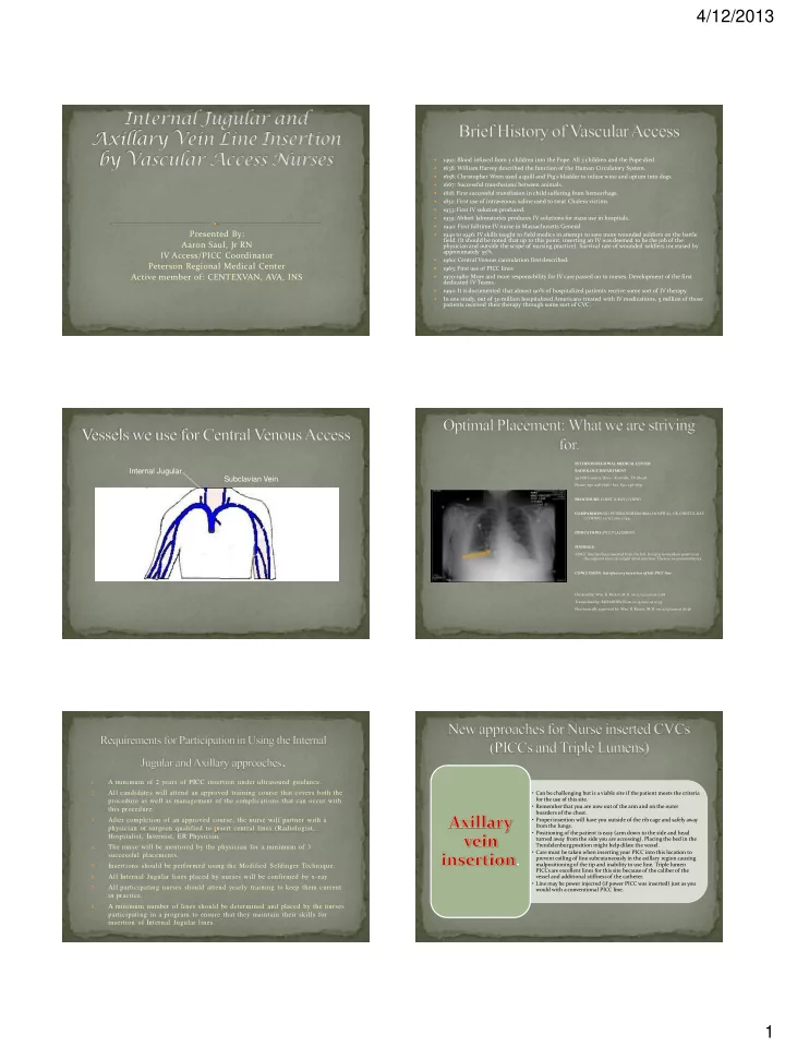
Presented By: Aaron Saul, Jr RN IV Access/PICC Coordinator Peterson Regional Medical Center Active member of: CENTEXVAN, AVA, INS

1492: Blood infused from 3 children into the Pope. All 3 children and the Pope died.

1638: William Harvey described the function of the Human Circulatory System.

1658: Christopher Wren used a quill and Pig’s bladder to infuse wine and opium into dogs.

1667: Successful transfusions between animals.

1818: First successful transfusion in child suffering from hemorrhage.

1832: First use of intravenous saline used to treat Cholera victims

1933: First IV solution produced.

1935: Abbott laboratories produces IV solutions for mass use in hospitals.

1940: First fulltime IV nurse in Massachusetts General.

1940 to 1946: IV skills taught to field medics in attempt to save more wounded soldiers on the battle

  • field. (It should be noted that up to this point, inserting an IV was deemed to be the job of the

physician and outside the scope of nursing practice). Survival rate of wounded soldiers increased by approximately 55%.

1960: Central Venous cannulation first described.

1965: First use of PICC lines

1970-1980: More and more responsibility for IV care passed on to nurses. Development of the first dedicated IV Teams.

1990: It is documented that almost 90% of hospitalized patients receive some sort of IV therapy.

In one study, out of 30 million hospitalized Americans treated with IV medications, 5 million of those patients received their therapy through some sort of CVC.

Internal Jugular External Jugular Subclavian Vein Axillary Vein Cephalic Vein Basilic Vein Brachial Vein

PETERSON REGIONAL MEDICAL CENTER RADIOLOGY DEPARTMENT 551 Hill Country Drive • Kerrville, TX 78028 Phone: 830-258-7636 • Fax: 830-258-7657 PROCEDURE: CHEST X-RAY (1 VIEW) COMPARISON: SID PETERSON MEMORIAL HOSPITAL, CR, CHEST X-RAY (2 VIEWS), 12/12/2010, 11:49. INDICATIONS: PICC PLACEMENT FINDINGS: A PICC line has been inserted from the left. Its tip is in excellent position at the superior vena cava/right atrial junction. There is no pneumothorax. CONCLUSION: Satisfactory insertion of left PICC line. Dictated by: Wm. R. Rector, M.D. on 12/13/2010 at 11:58 Transcribed by: RICHARDSON on 12/13/2010 at 12:43 Electronically approved by: Wm. R. Rector, M.D. on 12/13/2010 at 16:36

1.

A minimum of 2 years of PICC insertion under ultrasound guidance.

2.

All candidates will attend an approved training course that covers both the procedure as well as management of the complications that can occur with this procedure.

3.

After completion of an approved course, the nurse will partner with a physician or surgeon qualified to insert central lines (Radiologist, Hospitalist, Internist, ER Physician.

4.

The nurse will be mentored by the physician for a minimum of 3 successful placements.

5.

Insertions should be performed using the Modified Seldinger Technique.

6.

All Internal Jugular lines placed by nurses will be confirmed by x-ray.

7.

All participating nurses should attend yearly training to keep them current in practice.

8.

A minimum number of lines should be determined and placed by the nurses participating in a program to ensure that they maintain their skills for insertion of Internal Jugular lines.

  • Can be challenging but is a viable site if the patient meets the criteria

for the use of this site.

  • Remember that you are now out of the arm and on the outer

boarders of the chest.

  • Proper insertion will have you outside of the rib cage and safely away

from the lungs.

  • Positioning of the patient is easy (arm down to the side and head

turned away from the side you are accessing). Placing the bed in the Trendelenburg position might help dilate the vessel.

  • Care must be taken when inserting your PICC into this location to

prevent coiling of line subcutaneously in the axillary region causing malpositioning of the tip and inability to use line. Triple lumen PICCs are excellent lines for this site because of the caliber of the vessel and additional stiffness of the catheter.

  • Line may be power injected (if power PICC was inserted) just as you

would with a conventional PICC line.

.

slide-2
SLIDE 2

4/12/2013 2

1.

Thorough venous assessment with ultrasound of the patient should be performed on bilateral extremities to indeed identify that there are no usable vessels for traditional PICC insertion.

2.

Thorough history of any vascular access is very important as this will dictate which vessel (right or left) will be attempted.

3.

The patient’s chest mass must be taken into account. For example patient of my mass would not be a very viable candidate for an axillary PICC (without a harpoon).

4.

As with PICC lines, the presence of medical devices (i.e. pacemakers or history of a pacemaker) again should be considered because of the stenosis caused by insertion of the wires into the subclavian vein.

5.

It should be noted that this should not be your first stop if at all possible and your assessment of the patient should dictate how to best proceed.

6.

Once you have selected your site, vessel entry should be performed again using the Modified Seldinger Technique with a micorpuncture kit (this consists of using a 21 ga needle to access the vessel and wiring the vessel for purchase with a 0.018 wire).

7.

Obtain a chest xray to both confirm placement and ensure that there is no pneumothorax.

Axillary Vein Insertion Candidates:

Patients with a long history of numerous PICC insertions in both upper extremities and require venous access for treatment in a non-emergent situation.

Patients that are unable to position their arm for PICC

  • insertion. (Patients that have severe arthritis, rotator cuff

problems, shoulder replacements, other conditions).

Patients that require venous access but have no vessels in the upper extremities of suitable size for cannulation until reaching the level of the axillary vein.

Right Clavicle =Brachial Artery =Axillary Vein Left Clavicle =Brachial Artery =Axillary Vein Approximate boarder of the rib cage Lower boarder of the clavicle Axillary fold General area of axillary vein

slide-3
SLIDE 3

4/12/2013 3

Pectoral Muscle

  • This is a big jump in practice. Remember, it’s not if you have a

complication; IT’S WHEN.

  • Very intricate yet very simple site for insertion.
  • Again patient positioning is straight forward (head away from

side being accessed and Trendelenburg the bed).

  • With this site, air embolism is a VERY real danger, remember

what you need to do if this occurs (besides the expletive that will creep out under your breath and behind the mask).

  • These lines are excellent for the patient that requires CVP

monitoring in the ICU.

  • This is not a suitable site for the insertion of PICC lines.

(External jugular insertion of PICC lines is still being considered at this time and is actually being performed in some facilities across the country). 1.

Again, thorough venous assessment with ultrasound is of the utmost importance to identify the underlying structures at your proposed site of insertion. If separation of the Internal Jugular and Carotid is difficult or problematic, move to the other side to avoid inadvertently accessing the Carotid artery.

2.

The right Internal Jugular vein should be your first choice because it is a “straight shot” into the SVC to the Cavoatrial junction. The left Internal Jugular can be used but me mindful of the abrupt angle it takes when it joins the brachiocephalic vein crossing the

  • chest. (Accidental perforation can occur when dilating up to insert your catheter).

3.

Proper insertion should be as close to the base of the neck to ensure, in most cases, safe positioning of the Internal Jugular from the Carotid artery.

4.

Access is best achieved, if not emergent, by again using a micropuncture kit and the Modified Seldinger Technique so that if the Carotid artery is hit, you are not dealing with a 14 ga hole but instead with a 21 ga hole.

5.

If emergent, use great care with the 14 ga needle and perform the procedure as you would normally (MST).

6.

To help avoid the development of a hematoma, take the patient out of Trendelenberg as soon as you have the catheter in place.

7.

If possible, suture the catheter down to the collar bone ensuring that you have a gentle curve in the catheter.

8.

Again obtain a chest xray to both confirm placement and ensure that there is no pneumothorax.

slide-4
SLIDE 4

4/12/2013 4

1.

The patient has limited selections for venous access. Upper extremity co- morbidities preclude PICC placement.

2.

The patient needs access in a more emergent situation.

3.

Patients that need the most accurate CVP monitoring possible.

4.

Patients that will require large volumes of fluid and/or blood products (e.g. septic patients, GI bleeds, patients in cardiogenic shock (s/p cardiac arrest)).

5.

Patients with CKD.

6.

Patients with known coagulopathies may not be candidates for this

  • procedure. (Platelet count of less that 50,000 or an elevated INR).

7.

Any other patient that you think would benefit from have a large bore central line for treatment of their condition.

Boarder of the Sternocleidomastoid muscle Upper boarder of the Clavicle Approximate region of where the IJ and Carotid run

4.2% of time

12 o’clock 1 o’clock 2 o’clock 3 o’clock

16.4% of time 71.4% of time 9% of time

=Carotid Artery =Internal Jugular Vein

12 o’clock 11 o’clock 10 o’clock 9 o’clock

=Carotid Artery =Internal Jugular Vein 6.3% of time 75.5% of time 9% of time 9% of time

slide-5
SLIDE 5

4/12/2013 5

 Pneumothorax:

 Turn patient onto their right side to allow for full expansion of the left

lung and vice versa if approaching from the left IJ.

 Notify the primary care physician immediately so that further

interventions can be instituted.

 Monitor: respiratory effort, oxygen saturation, blood pressure, heart rate,

and level of consciousness.

 In severe cases, call a Code to get a physician to the bedside

  • immediately. Emergency needle decompression can be performed until

the physician arrives to place a chest tube.

 This procedure is done by inserting a 14 gauge IV catheter between the first

and second rib at the midclavicular line. Using the finger cut from a glove (both ends open) secure one end to the catheter with a rubber band or tap so that it acts as a Heimlich valve.

 Hemothorax:

 Most often a result of a “through and through” arterial stick

while attempting access. (This is much more common in your patients that have coagulopathies).

 Again, immediately notify the primary care physician and if

necessary, call a Code to get a physician immediately to the bedside.

 Monitor blood pressure, heart rate, respiratory effort, and

level of consciousness.

 Chest tube placement must be performed in order to

evacuate the blood and if severe enough an emergency thoracotomy to stop the bleeding.  Air Embolism:

 This event requires that the patient be placed immediately

  • n the Left side with the bed in Trendelenberg position to

prevent the “air bubble” from entering the region of the pulmonic valve in the right ventricle.

 Again closely monitor the patients blood pressure,

respiratory effort, level of consciousness, and administer 100% O2.

 Notify the primary care physician immediately for orders on

how to proceed.

 If the catheter tip is in the region of the right atrium, an

attempt can be made to aspirate the air (this should be done at the discretion of the clinician inserting the line and/or the physician).

slide-6
SLIDE 6

4/12/2013 6

 Vessel Access, Cannulation, and Line Removal:

 If the patient presents with sclerotic vasculature and as the clinician you have

difficulty accessing the vessel, the patient should be referred to the Interventional Radiology department and the ordering physician notified. This is why it is very important to perform a thorough assessment of your patient with ultrasound and familiarizing yourself with the patient’s medical history before proceeding.

 If you are unable to advance and/or retract your wire then the procedure should

be halted and again notify the ordering physician for further direction.

 Difficulty in dilating the tissue for line insertion or inadvertent vessel

perforation with the dilator are complications that can be encountered.

 If resistance is encountered during removal of a device, then again the procedure

should be halted to prevent damage to the vessel and harm to the patient.

 When in doubt, PUNT.

Axillary PICC

You are using the same kit that you already have for conventional PICC insertions. In most cases, there is no increase in cost (with the average cost

  • f a full barrier kit running around $180) of doing

business.

Your site of insertion is not on the arm but on the chest just under the shoulder.

You are performing the same technique for vessel access as with a conventional PICC line.

The risk for Pneumothorax is a much more real complication vs. insertion of a conventional PICC.

In most cases, it is a much easier insertion that with a conventional PICC.

Procedural time is 30-45 minutes.

You still have the benefit of a low infection rate due to the location of the insertion site.

Kit price is usually less to just slightly more than your PICC kit (if using a full barrier kit) of $170- $180.

More risk involved in procedure because of site

  • f insertion.

Again, you are performing the same procedure to access the vessel (e.g. MST).

Again, complications of the procedure (e.g. Pneumothorax, Arterial Puncture, and Air Embolism) are high and should be taken into consideration when selecting to insert a line at this site.

Procedural time, once you have your rhythm, is also 30-45 minutes.

Internal Jugular Line

 Infection rates for Internal Jugular lines catheters averages 22%

per 1000 catheter days or 113 lines out of 515 lines placed in a study published in the European Journal of Microbiology and Infectious Diseases.

 The most common organism causing infections being a Gram-

positive bacteria (Coagulase-negative staphylococci).

 Another study (Lippincott’s Nursing Center

www.nursingcenter.com) stated that for PICC’s, the

infection rate was 2.4% or 2.1 episodes per 1000 catheter days compared to CVC rates of 4.4% or 2.7 episodes per 1000 catheter days.

 Sample Note:

Order received and verified for Central line placement. Risks, benefits, and alternative treatments/options discussed with the patient and signature on consent

  • witnessed. Patient’s medical records, to include allergies, were reviewed. Time out

called and patient identifiers verified. ## neck was then prepped with ########## and draped in the usual sterile manner. # ml of 1% lidocaine was used as a local

  • anesthetic. With the use of ultrasound guidance, the vessels were assessed, imaged

and determined patent. The ## internal jugular vein was accessed using the modified Seldinger technique on the # attempt. Blood return was non-pulsatile and venous in

  • color. An S/D/T lumen Central line was then inserted and flushed with ## ml

normal saline per port. The Central line was then secured with 3-0 Ethilon sutures and covered with a Tegaderm with CHG gel dressing. Stat chest x-ray ordered for tip placement. Charge/Primary nurse instructed to not use line until tip confirmation

  • btained. EBL <# ml. Tip of catheter in P/M/D SVC on chest xray. Charge/Primary

nurse notified of tip position and given clearance to use line at this time.

Questions??