
SLIDE 1
12/21/2012 1
Hip Labral Pathology – From Diagnosis to Functional Rehabilitation
Josette Fisher, PT, ATC, CSCS Director of Rehabilitation Jfisher@excelsiorortho.com
Objective
- Overview of labral tears
- Hip impingement
‐what does that mean?
- Review of traditional exam
- Treatment philosophy
- How functional assessment can confirm diagnosis and drive treatment
plan
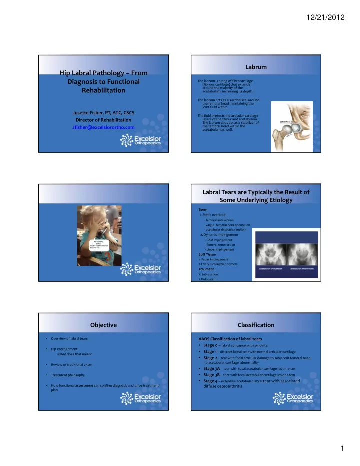
Labrum
The labrum is a ring of fibrocartilage (fibrous cartilage) that extends around the majority of the acetabulum, increasing its depth. The labrum acts as a suction seal around the femoral head maintaining the joint fluid within. The fluid protects the articular cartilage layers of the femur and acetabulum. The labrum does act as a stabilizer of the femoral head within the acetabulum as well.
Labral Tears are Typically the Result of Some Underlying Etiology
Bony
- 1. Static overload
‐ femoral anteversion
‐ valgus femoral neck orientation ‐ acetabular dysplasia (ant/lat)
- 2. Dynamic Impingement
‐ CAM impingement ‐ femoral retroversion ‐ pincer impingement
Soft Tissue
- 1. Psoas Impingement
2.Laxity – collagen disorders
Traumatic
- 1. Subluxation
2.Dislocation
AAOS Classification of labral tears
- Stage 0 – labral contusion with synovitis
- Stage 1 – discreet labral tear with normal articular cartilage
- Stage 2 – tear with focal articular damage to subjacent femoral head,
no acetabular cartilage abnormality
- Stage 3A – tear with focal acetabular cartilage lesion <1cm
- Stage 3B – tear with focal acetabular cartilage lesion >1cm
- Stage 4 – extensive acetabular labral tear with associated