1 IG / TCR heterogeneity : combinatorial and junctional diversity - - PDF document

1
SMART_READER_LITE
LIVE PREVIEW

1 IG / TCR heterogeneity : combinatorial and junctional diversity - - PDF document

Lymphoid malignancies: leukemias and lymphomas Red blood cell Platelet Granulocyte Molecular diagnostics of Monocyte lymphoid malignancies Myeloid lymphoma stem cell leukemia Chronic Acute B-lymphocyte Stem cell 11 th Course on


slide-1
SLIDE 1

1

Molecular diagnostics of lymphoid malignancies

11th Course on Molecular Diagnostics Molecular Medicine Postgraduate School, Erasmus MC, November 3, 2017

  • Dr. A.W. Langerak (Ton)

Laboratory for Medical Immunology, Dept. Immunology Erasmus MC, Rotterdam a.langerak@erasmusmc.nl

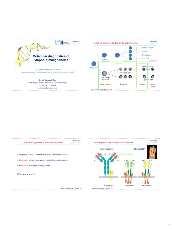
Stem cell Myeloid stem cell B-lymphocyte T-lymphocyte Precursor cells Red blood cell Platelet Granulocyte Monocyte

Bone marrow Thymus Blood

Lymphoid stem cell

Lymphoid malignancies: leukemias and lymphomas

  • Dept. of Immunology, Erasmus MC

Lymph node lymphoma leukemia Acute Chronic

 Diagnosis : tumor (= clone of cells) or e.g. immune response ?  Prognosis : (clinical) heterogeneity and differences in outcome  Monitoring : evaluation of therapy effect What markers to use ?

  • Dept. of Immunology, Erasmus MC

Molecular diagnostics in leukemia / lymphoma

IgH IgL IgL

V V C C J J

IgH

V V D D J J C C C C C C C C C D 7 9 a C D 7 9 b

CD3 e CD3 d CD3 g CD3 x CD3 x

V J C V D J C

TCR a TCR b

B-lymphocyte T-lymphocyte

CD3 e CD3 d CD3 g CD3 x CD3 x

V J C V D J C

TCR g TCR d

T-lymphocyte

  • Dept. of Immunology, Erasmus MC

immunoglobulin T-cell receptor Immunoglobulin and T-cell receptor molecules Antigen binding

CDR’s

slide-2
SLIDE 2

2

  • Dept. of Immunology, Erasmus MC

1 2 3 4 5 6 66 1 2 3 4 1 2 3 4 5 6

V

H

D

H

J

H

C m s DJ joining VD-J joining precursor IGH mRNA mature IGH mRNA transcription RNA splicing V D J C m

27

IgH IgL IgL

V V C C J J

IgH

V V D D J J C C C C C C C C C D 7 9 a C D 7 9 b

translation

IG / TCR heterogeneity : combinatorial and junctional diversity

  • Dept. of Immunology, Erasmus MC

GTATTACGATTTTTGGAGTGGTTATTATACC IGHV3-21 IGHJ4-1 IGHCM IGHD3-3

insertion insertion IGHV3-21 (germline)

TGTATTACTGTGCGAGA

IGHD3-3 (germline) IGHJ4-1 (germline)

ACTACTTTGACTACT TGTATTACTGT TGTATTACTGTGCG TGTATTACTGTGC TGTATTACTGTGCGAG TGTATTACTG TGTATTACTGTGCG TGTATTACTGTGCGA TGTATTACT TGTATTACTGTGCG TGTATTACTGTGC TGTATTACTGTGCGAGA TGTATTACTGTG TGTATTA TGTATTACTGTGC TGTATTACTGTGCGA CGATTTTTGGAGTGGTTATTATA TTA CGATTTTTGGAGTGGTTATTATAC TTTTGGAGTGGTTATTATACC TATTACGATTTTTGGAGTGGTTAT CGATTTTTGGAGTGGTTATTATA TACGATTTTTGGAGTGGTTATTAT TTACGATTTTTGGAGTGGTTATTATACC GATTTTTGGAGTGGT ATTACGATTTTTGGAGTGGTTATTATA TTTGGAGTGGTTATTATA ATTACGATTTTTGGAGTGGTTATTATACC TATTACG ATTTTTGGAGTG CGATTTTTGGAGTGGTTATTATA ATTACGATTTTTGGAGTGG GTATTACGATTTTTGGAGTGGTTATTATACC TGACTACT CTTTGACTACT ACTACTTTGACTACT TTTGACTACT ACTTTGACTACT TGACTACT TACTTTGACTACT ACTACTTTGACTACT CTTTGACTACT ACTTTGACTACT GACTACT CTACTTTGACTACT ACT TTTGACTACT ACTTTGACTACT

AGGC TATCCGGA CCGGACTG CTGAGTC ACATCGA CGT CCGG GATG TTCA GGCTAG GTCCAG CCGGA ACGC CGTA GTCCA CGATCG GGT CGTAGCGTA CGTAG GGCTAAGG CGGAGC GGTTC CGATCGA CC GTCG CCGTAG C GTACG GGCA

junctional region

IG / TCR heterogeneity : junctional diversity

4 3 6 ++++ ++   13 2 > 70 5 4

  • > 50

> 40 TCRd TCRb Ig Ig > 1012 > 1012 > 1012 Estimation of total primary receptor repertoire ++ + ++ Junctional region diversity > 5000 > 5 x 10⁶ > 5 x 10⁶ Combinatorial diversity 5 55 6

  • J genes
  • 27
  • D genes

6 > 50 > 100

  • V genes

Number of genes TCRg TCRa IgH TCRgd TCRab

  • Dept. of Immunology, Erasmus MC

Estimated potential primary repertoire of human IG / TCR molecules

B-lymphocyte T-lymphocyte Precursor cells

Bone marrow Thymus Blood

Lymphoid stem cell

  • Dept. of Immunology, Erasmus MC

Lymph node Ig genes rearranged TCR genes rearranged  normal B/T cells: unique IG / TCR gene rearrangement  lymphoid tumor: all cells will have identical Ig or TCR genes (monoclonal) IG / TCR rearrangements in leukemia and lymphoma

slide-3
SLIDE 3

3

 Diagnosis : tumor (= clone of cells) or e.g. immune response ? Clonality testing using IG / TCR rearrangements, as polymorphic markers  PCR-based fragment analysis / spectratyping

  • Dept. of Immunology, Erasmus MC

Molecular diagnostics in leukemia / lymphoma

  • non-hemopoietic tissue : Ig / TCR segments far apart  no PCR products
  • normal BM, PB, lymph node : Ig / TCR segments coupled  PCR products

further discrimination between polyclonality and monoclonality:  GeneScan fragment analysis : length of PCR products

  • Dept. of Immunology, Erasmus MC

1 2 3 4 5 6 66 1 2 3 4 1 2 3 4 5 6

V

H

D

H

J

H

C m s DH  JH joining VH  DH-JH joining

27

PCR-based analysis of Ig / TCR rearrangements

DNA fragment length fluorescence intensity

Clonality testing  CDR3 heterogeneity

  • Dept. of Immunology, Erasmus MC

DNA fragment length dominant peak = clone fluorescence intensity

Clonality testing  CDR3 heterogeneity

  • Dept. of Immunology, Erasmus MC
slide-4
SLIDE 4

4

IGH gene analysis : multiplexing  7 IGHV families

Leukemia 2003;17:2257-2317

IGH gene analysis : multiplexing

>100 27 6

  • FR1

67 %

  • FR1

73 %

  • FR2

57 %

  • FR2

76 %

  • FR3

47 %

  • FR3

52 %

  • all FR

77 %

  • all FR

84 % DLBCL (n=116) FCL (n=109)

Leukemia 2007;21:207-214

DLBCL: Diffuse large B-cell lymphoma; FCL: Follicular Cell Lymphoma

B-cell lymphoma Complementarity of IG targets

T T T T T DC DC DC T T T T T T

naive (virgin) B-cells memory B-cells plasma cells IGH class switch recombination (CSR) positive selection

  • f B-cells for

antigen binding B-cell proliferations and somatic hypermutation (SHM)

Lymph node follicle

  • Dept. of Immunology, Erasmus MC

Somatic hypermutation can hamper primer annealing

GGATTCACCTTCAGTAGCTATAGC GGATTCACCTTCAGTAGCTATAGC GGATTCACCTTCAGTAGCTATAGC GGATTCACCTTCAGTAGCTATAGC GGATTCAC TTCAGTAGCTATAGC GGATTCACCTTCAGTAGCTATAGC GGATTCACCTTCAGTAG TATAGC GGATTCACCTTCAGTAGCTATA C GGATTCACCTTCAGTAGCTATAGC G T A VH3-21 JH4-1 DH3-3 CDR1 FR1 TCCTGTGCAGCCTCT FR2 ATGAACTGGGTCCGCCAGGCTCCAGGGAAG ATGAACTGGGTCCGCCAGGCTCCAGGGAAG ATGAACTGGGTCCGCCAGGCTCCAGGGAAG ATGAACTGGGTCCGCCAGGCTCCAGGG AG ATGAACTGGGTCCGCCAGGC CCAGGGAAG ATGAACTGGGTCCGCCAGGCTCCAGGGAAG ATGAACTGGGTCCGCC GGCTCCAGGGAAG ATGAA TGGGTCCGCCAGGCTCCAGGGAAG ATGAACTGGGTCCGCCAGGCTCCAGGGAAG G A G A CDR1 CDR3 CDR2 TCCTGTG AGCCTCT TCCTGTGCAGCCTCT TCCTGTGCAGCCTCT TCCTGTGCAGCCTCT TCCTGTGCAGCCTCT TCCTGTGCAGCCTCT TCCTGTGCAGCCTCT TCCTGTGCAGCCTCT T GGATTCACCTTCAGTAGCTAT GC GGATTCACCTTCAGTAG ATA G TA CT ATGAACTGGGTCCGCCAGGCTCCAGGGAAG ATGAACTGGGTCCGCCAGGCTCCAGGGAAG TCCTGTGCAGCCTCT TCCTGTGCAGCCTCT SHM:

selection for B-lymphocytes with high-affinity antibodies FR1 FR2 FR3

 Physiology: antibody affinity maturation  Diagnostics: primer misannealing

antibodies secondary responses bind antigen clearance

slide-5
SLIDE 5

5

  • FR1

67 %

  • FR1

73 %

  • FR2

57 %

  • FR2

76 %

  • FR3

47 %

  • FR3

52 %

  • all FR

77 %

  • all FR

84 %

  • all FR + DH-JH

83 %

  • all FR + DH-JH

86 %

  • IGH + IGK

91 %

  • IGH + IGK

100 % DLBCL (n=116) FCL (n=109)

Leukemia 2007;21:207-214

DLBCL: Diffuse large B-cell lymphoma; FCL: Follicular Cell Lymphoma

B-cell lymphoma  Clonality testing possible in vast majority of patients Complementarity of IG targets

 Prognosis : (clinical) heterogeneity and differences in outcome Somatic mutation analysis of rearranged IG genes  Sequencing

Alternative targets:

  • chromosome aberrations / translocations / deletions
  • mutations / SNV’s
  • Dept. of Immunology, Erasmus MC

Molecular diagnostics in leukemia / lymphoma

  • Tumor of mature circulating B-cells
  • High leukocyte counts
  • Monomorphic small round lymphocytes

Clinical features

  • Most patients asymptomatic
  • Others show fatigue, splenomegaly, lymphadenopathy
  • Clinical course mostly indolent, but can also be more aggressive

 search for prognostic factors !!

  • Dept. of Immunology, Erasmus MC

~1995 : somatic hypermutation (SHM) status of IGHV gene: unmutated: poor prognosis mutated: favorable prognosis B-cell chronic lymphocytic leukemia (CLL)

  • IGH FR1 BIOMED-2 multiplex PCR + sequencing
  • blast in IMGT database
  • determine V, D, J gene usage, reading frame

IGHV mutational analysis

slide-6
SLIDE 6

6

98-100% homology to closest IGHV = non-mutated <98% homology to closest IGHV = mutated

IGHV mutational analysis

  • determine total number of mutations
  • calculate % homology : # mutations / # bases in V gene

60 Overall survival mutation (%germ < 98) Cumulative percentage 12 24 36 48 60 25 50 75 100 no yes months

At risk: no 52 26 16 7 3 y es 82 54 27 10 6 Logrank P<.001 no 52 17 y es 82 6 N F

Haematologica 2006;91:56-63

Erasmus MC CLL cohort Survival according to IGHV mutational status Stereotypy of Ig receptors and clinical impact

Ghia et al, Blood 2005 Tobin et al., Blood 2002, 2003 IG homology in various CLL patients:

 Antigen-driven pathogenesis?

 Monitoring : evaluation of therapy effect Minimal residual disease analysis using patient-specific IG/TCR rearrangements  real-time quantitative PCR (RQ-PCR)

  • Dept. of Immunology, Erasmus MC

Molecular diagnostics in leukemia / lymphoma

slide-7
SLIDE 7

7

  • Dept. of Immunology, Erasmus MC

B-lymphocyte T-lymphocyte Precursor cells

Bone marrow Thymus Blood

Lymphoid stem cell

Lymph node 85% 15% Acute lymphoblastic leukemia (ALL) ALL is most common form of cancer in childhood

  • Prognosis: after 5 years ~90% still alive
  • Many short-term and long-term side effects
  • Recognition of poor risk and high risk patients is important in order to adjust therapy

Characteristics of tumor cells

e.g.

  • chromosomal translocations
  • gene expression profile
  • immunophenotype
  • in vitro drug sensitivity

In vivo drug distribution

e.g.

  • gastrointestinal absorption
  • distribution in body (e.g. CNS)
  • drug metabolism (e.g. polymorphisms in enzymes)
  • liver excretion
  • kidney excretion

Treatment compliance

e.g.

  • duration of Rx
  • side effects (e.g. allergy, infections)

Evaluation of overall treatment effectiveness by MRD diagnostics

1 2 3 4 r e l a t i v e f r e q u e n c y

  • f

l e u k e m i c c e l l s follow-up in years “cure” 1 10

  • 1

10

  • 2

10

  • 3

10

  • 5

10

  • 4

10

  • 6

10

  • 7

I C II Maintenance Rx

  • Dept. of Immunology, Erasmus MC

Treatment effectiveness in childhood ALL

20 40 60 80 20 40 60 80 100 Low-risk group (n=50; 46%) p(trend)<0.001 intermediate-risk group (n=47; 43%) high-risk group (n=12; 11%) MRD >10-3 at TP1 and TP2 MRD negative at TP1 and TP2

% Relapse-free survival

Lancet, 1998; Blood 2002

months from time point 2

 MRD is a strong and independent prognostic factor  Sensitive (10-4) and quantitative data are required: RQ-PCR Survival of BCP-ALL according to MRD-based risk groups

  • A. Identification of targets at diagnosis (PCR analysis, sequencing)

 in >95% of patients at least two Ig/TCR markers ( sequencing)

  • B. Set-up and testing of patient-specific RQ-PCR assays
  • Design of patient-specific RQ-PCR assay
  • Sensitivity testing
  • C. Analysis of MRD during follow-up using RQ-PCR analysis
  • MRD analysis of follow-up samples
  • Control gene analysis of follow-up samples
  • Dept. of Immunology, Erasmus MC

VH3-21 DH3-3

MRD monitoring via Ig/ TCR markers

slide-8
SLIDE 8

8

  • 1/CTGAGTC/-1

Patient-specific primer

VH3-21 JH4 DH3-3

  • 6/CGTAGCGTA/-5

Common probe Common reverse primer

CDR3 fingerpint: patient-specific RQ-PCR design

  • Dept. of Immunology, Erasmus MC

Junctional region = DNA fingerprint of leukemic cell

<10 <3.5 No 3’ hairpin

1.E-04 1.E-03 1.E-02 1.E-01 1.E+00 1.E+01 10 20 30 40 50 Cycle delta Rn (‘fluorescence’) 10 = 10% 10 = 1% 10 = 0.1% 10 = 0.01% 10 = 0.001% MNC (6-fold)

  • 1
  • 2
  • 3
  • 4
  • 5

threshold

  • Dept. of Immunology, Erasmus MC

Standard curve: dilutions of diagnostic sample

  • A. Identification of targets at diagnosis (PCR analysis, sequencing)
  • B. Set-up and testing of patient-specific RQ-PCR assays
  • Design of patient-specific RQ-PCR assay
  • Sensitivity testing
  • C. Analysis of MRD during follow-up using RQ-PCR analysis
  • MRD analysis of follow-up samples
  • Control gene analysis of follow-up samples
  • Dept. of Immunology, Erasmus MC

VH3-21 DH3-3

MRD monitoring via Ig/ TCR markers

slide-9
SLIDE 9

9

10

  • 5

30 25 20 35 40

c y c l e t h r e s h

  • l

d ( C T ) dilution

10

  • 4

10

  • 3

10

  • 2

10

  • 1

standard curve (dilutions of diagnostic sample)

Slope: -3.3

2 (21) - fold dilution  + 1 Ct 4 (22) - fold dilution  + 2 Ct 8 (23) -fold dilution  + 3 Ct 10 (23.3) -fold dilution  + 3.3 Ct

  • Dept. of Immunology, Erasmus MC

MRD level analysis  Data analysis according to international guidelines (EuroMRD)

1.E-04 1.E-03 1.E-02 1.E-01 1.E+00 1.E+01

10 20 30 40 50 Cycle delta Rn 10 10 10 10 10 MNC

  • 1
  • 2
  • 3
  • 4
  • 5

Quantitative range Sensitivity

quantified positive negative Leukemia 2008, Leukemia 2007

MRD data interpretation

50 100

weeks n

  • r

m a l i z e d M R D l e v e l

150 10

  • 6

10

  • 5

10

  • 4

10

  • 3

10

  • 2

10

  • 1

1 precursor-B-ALL DCLSG 5257 IGK PCR target (10-5 ) I clinical phase D C I maintenance-Rx CR R

  • Dept. of Immunology, Erasmus MC

Current MRD diagnostics

50 100

weeks n

  • r

m a l i z e d M R D l e v e l

150 10

  • 6

10

  • 5

10

  • 4

10

  • 3

10

  • 2

10

  • 1

1 precursor-B-ALL DCLSG 5257 IGK PCR target (10-5 ) I clinical phase D C I maintenance-Rx CR R

High Risk

 More intense therapy in current ALL protocol

?

  • Dept. of Immunology, Erasmus MC

Current MRD diagnostics

slide-10
SLIDE 10

10

Diagnosis IG/TCR clonality testing (multiplex PCR) : important and reliable tool for diagnosing esp. mature leukemias and lymphomas Prognosis Classification of CLL on basis of IGHV SHM status (via sequencing) : prognostic + potentially therapeutic implications Monitoring Detection of MRD in childhood ALL by IG / TCR ASO RQ-PCR: therapy stratification

  • Dept. of Immunology, Erasmus MC

TAKE HOME MESSAGES Mol. diagnostics in lymphoid malignancies

What is the future of molecular diagnostics in lymphoid leukemia ???

IG-TCR : low- vs. high-throughput cell population low-throughput high-throughput

Langerak, J Immunol 2017

Pros of NGS immunogenetics

Clonality

  • clonal relationship
  • intra-clonal diversity

MRD

  • target identification;
  • sensitive monitoring
  • subclonal heterogeneity + clonal evolution

Repertoire (normal, clonal)

  • depth and coverage
slide-11
SLIDE 11

11

Immunogenetics

IG / TR rearrangements as unique “molecular signatures” with implications for

  • research

B / T cell ontogeny, immunodeficiency, autoimmunity, lymphomagenesis

  • clinical diagnostics

clonality assessment MRD assessment repertoire analysis

 EuroClonality-NGS main objective : develop, standardize, and validate IG/TR NGS assays for clinical applications

NGS clonality: mono- vs. polyclonal (IGK)

Sample PBMC Sampl: lymphoma

feature type A: junction aa length feature type B: clonotype feature type A: junction aa length feature type B: clonotype Software: ARResT / Interrogate

NGS clonality: clonal relationship

Sampl: lymphoma year 1 and 2

feature type A: junction aa length feature type B: clonotype feature type A: junction aa length feature type B: clonotype Software: ARResT / Interrogate

Detection of clonal evolution

STUDY BACKGROUND

IG/TR NGS-based MRD Workflow Screening

  • f diagnostic sample

MRD-Analysis

Preparation of amplicons for HTS HTS Bioinformatic identification of index-sequence IG/TR Multiplex PCR Preparation of amplicons for HTS HTS Bioinformatic search for index- sequence IG/TR Multiplex PCR Courtesy: M. Brueggemann

No need for clone specific assay Increased sensitivity Increased specificity Detection of

  • ligoclonality

Insights into background B- and T- cell repertoire

Promises of HTS

slide-12
SLIDE 12

12

Challenges of NGS immunogenetics

Multiplexing

Limits of detection (quantifiable, sensitivity) Coverage Equal representation

Error correction

Standardization

Validation

Redefinition of clonality

Data visualization

Proof-of-principle studies showing potential for different diagnostic applications Routine diagnostic applications requires further standardization and clinical validation (ongoing in European consortium) NGS will quickly take over current methods for clonality analysis, MRD target identification and monitoring, and deep immune repertoire analysis

  • Dept. of Immunology, Erasmus MC

SUMMARY NGS-based diagnostics in leukemia / lymphoma

Past & present members

  • f the Langerak lab

Erasmus MC