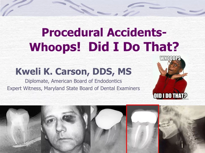
SLIDE 1 Procedural Accidents- Whoops! Did I Do That?
Kweli K. Carson, DDS, MS
Diplomate, American Board of Endodontics Expert Witness, Maryland State Board of Dental Examiners
SLIDE 2
Types of Procedural Accidents
Crown and root perforations Iatrogenic canal obstruction (due to dentin shavings or dental materials) Ledge formation Separated instruments Swallowed or aspirated instruments Sodium hypochlorite incidents Underfilled or overfilled root canals Post space perforations Vertically fractured roots Treating the wrong tooth
SLIDE 3
Preventing Procedural Accidents
Thorough clinical and radiographic exam Accurate diagnosis and treatment planning Use the AAE Endodontic Case Difficulty Assessment Form and Guidelines to determine risk factors Refer potentially difficult or challenging cases to an endodontist
SLIDE 4
AAE Endodontic Case Difficulty Assessment Form and Guidelines
SLIDE 5
Procedural Accidents Occur
During Access Preparation During Cleaning and Shaping During Obturation During Post Space Preparation and Post Insertion
SLIDE 6 Procedural Accidents During Access Preparation
Perforations
Coronal - lateral Coronal - furcation
Iatrogenic canal
SLIDE 7
Perforations
Iatrogenic perforation
Artificial opening in a tooth created by drilling, boring, piercing, or cutting through tooth structure (with burs, hand instruments) Communication between pulp system and external tooth surface
SLIDE 8
Perforation During Access Preparation
SLIDE 9 Prevention During Access
Clinical examination
Rotation, tipping of crown Cast crown may not represent
Radiographic examination
SLIDE 10
Prevention During Access
Orientation of bur
Depth of pulp chamber roof Height of pulp chamber Location of pulp horns Level of chamber floor
Be Patient ENDO-Z bur (non-cutting tip)
SLIDE 11
Recognition of Perforations During Access
Sudden appearance of persistent hemorrhage Radiographic extrusion of a file into PDL or bone.
SLIDE 12
Identification of Perforations During Access
Dental operating microscope Electronic apex locator
SLIDE 13 Treatment of Perforations
Lateral perforations can be repaired with geristore, composite or a crown (microscope recommended).
Al-Sabek, F. et al JOE 2005: Vol 31, Num. 3, pp 205-208 “In vitro interpretation indicates that Geristore is less cytotoxic to gingival fibroblasts [than Ketac-Fil or IRM]”
SLIDE 14
Treatment of Perforations
Furcal perforations can be repaired with MTA or amalgam (microscope recommended) – a file or paper point should be placed in the canals to prevent blockage of canals with the repair materials.
SLIDE 15 Perforation Material - MTA
Nakata, T. et al JOE 1998: Vol 23, Num. 4, pp 184-196 “MTA was significantly better than amalgam at preventing bacterial leakage in furcal perforation repairs.”
Mineral Trioxide Aggregate
SLIDE 16 Factors Affecting Prognosis – Perforations
Relationship of perforation to the gingival sulcus Time lapse before perforation repair Adequacy of perforation repair Sterility of perforation repair Material used to seal perforation
Johnson W. et al JADA 1988: Vol 117, Num. 3, pp 473-476
SLIDE 17
Iatrogenic Canal Obstructions
Restorative materials and dentin shavings can travel into canals during access
SLIDE 18
Iatrogenic Canal Obstructions
Prevention
All necessary restorative material should be removed prior to exposing the pulp Place a small cotton pellet over the canal orifices if access is enlarged Frequent and copious irrigation Place files or paper points in canals if using repair materials
SLIDE 19
Procedural Accidents During Cleaning and Shaping
Ledge formation Root perforations (apical, lateral, coronal) Separated instruments Swallowing or aspirating instruments Extrusion of irrigants into periapical tissues
SLIDE 20 Ledge Formation
Created when the working length (WL) can no longer be negotiated Causes – inadequate straight-line access, filing a curved canal short of WL, over- enlargement of a small curved canal, debris packed in apical canal area
NORMAL
SLIDE 21 Ledge Formation
“Canal curvature was the most significant variable affecting the incidence of ledging”
Kapalas A. et al Endod Dent Traumatal 2000: Vol 16, Num. 5, pp 229-231
SLIDE 22
Preventing Ledging During Cleaning & Shaping
Straight line access Use small flexible files especially with curved canals (pre-curve if nec) Avoid forcing large files into curved canals Estimated working length verified by apex locator or radiograph
SLIDE 23
Root Perforations During C&S (apical, lateral, coronal)
APICAL LATERAL CORONAL
SLIDE 24
Identification of Root Perforation
Observation of bleeding
Direct Indirect (paper points)
Radiographs
Small file
Apex Locator
SLIDE 25 Management of Root Perforations
Treatment plan depends on
Accessibility Visibility Perforation size Periodontal conditions Strategic importance of tooth Patient’s oral hygiene Quality of root canal treatment Experience of the operator
Alhadainy, HA Oral Surg Oral Med Oral Path 1194: Vol 78, Num. 3, pp 368-374
SLIDE 26 Repair Materials
MTA
Less leakage in lateral root perforations than amalgam or IRM
Lee, 1993
SLIDE 27
Root Repair Materials
SLIDE 28
Root Perforation (mesio-buccal) - Apicoectomy
SLIDE 29 Root Perforations
Iatrogenic perforation of a root surface during endodontic treatment or restorative procedures may:
Decrease prognosis Cause secondary periodontal involvement Cause the loss of the tooth
Alhadainy, HA Oral Surg Oral Med Oral Path 1994: Vol 78, Num. 3, pp 368-374
SLIDE 30 Separated Instruments
Potential hazard that patients should be informed of prior to treatment Usually occur in small, long, curved, calcified or irregular canals
“Factors attributed to breakage of rotary files include canal curvature and other anatomic challenges, practitioner experience, frequency of use, and speed of rotation” Fishelburg G. et al Compend Contin Educ Dent 2004: Vol 25, Num 1, pp 17-24
SLIDE 31
How to Minimize Separation
Straight line access Light touch – gentle pressure Never force a file – take to resistance Consistent RPM
SLIDE 32
How to Minimize Separation
Use plenty of irrigation/lubrication Establish finger rest – minimizes pulling into canal Instrument with rotaries to point of divergence or trouble spots
SLIDE 33
How to Minimize Separation
File turning upon entry Know exact WL Clean and examine files frequently “When in doubt – throw it out”
SLIDE 34
Treatment – Separated Instruments
Depends on location Ultrasonics to remove Bypass Leave in place (guarded prognosis – possible apicoectomy in the future)
SLIDE 35 If there is ONE thing you remember about endodontics from this lecture, remember this…
ALWAYS use a rubber dam!!
Swallowing or Aspirating Instruments
SLIDE 36 Swallowing or Aspirating Instruments
“The use of rubber dam is an absolute essential during endodontic treatment”
Lambrianidis T. et al Endod Dent Traumatol 1996: Vol 12, Num 6, pp 301-304
“The placement of a rubber dam is considered the standard of care”
Fishelburg G. et al JOE 2003: Vol 29, Num 3, pp 683-684
SLIDE 37
ALWAYS USE A RUBBER DAM!
SLIDE 38
Extrusion of Irrigants into Periapical Tissues
Prevention – use side-slotted needle, keep needle moving, do not wedge needle into canal Recognition – prolonged severe pain followed by rapid diffuse swelling Treatment – reassurance, patient education, analgesics, multiple follow-up visits
SLIDE 39
Procedural Accidents During Obturation
Underfilling Overfilling Vertical fracture (rare)
SLIDE 40
Underfilling
Causes
Natural barrier Ledge formation Insufficient flaring No straight-line access Poorly adapted master cone Inadequate condensation pressure
SLIDE 41
Underfilling
Treatment
Depends on several factors
Radiographic findings Post present Crown margins
Retreatment Surgery (apicoectomy)
SLIDE 42
Overfilling
Extruded material can cause tissue damage and inflammation Caused by overinstrumentation through the apical foramen If overfilling is suspected, do working radiograph prior to searing gutta percha (remove all gutta percha if necessary) Treatment – mainly surgery (apicoectomy)
SLIDE 43
Vertical Fracture
Can be caused by condensation forces during obturation (rare)
SLIDE 44
Procedural Accidents During Post Space Preparation
Misdirected post preparation Perforation Prevention
Examine radiograph carefully Use heat to remove coronal gutta percha – to guide post drill
SLIDE 45
Procedural Accidents During Post Insertion
Use of an excessively large post, leading to vertical root fracture
SLIDE 46
Treating the Wrong Tooth
SLIDE 47
Discussing Procedural Accidents With Patients
Be honest / Look them in the eye Review your informed consent form Inform them about the accident Tell them how prognosis is affected Discuss procedures necessary for correction Consider referring the patient Help them financially (depends on the issue) Call your malpractice carrier for advice
SLIDE 48
Questions?
SLIDE 49
Thank You!