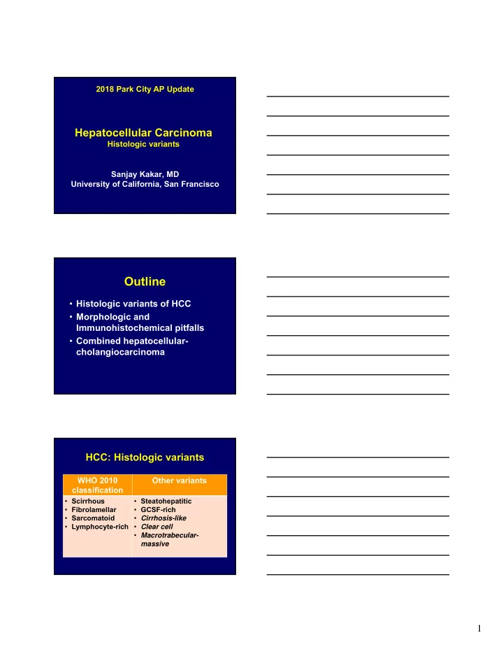
SLIDE 1 1
Hepatocellular Carcinoma
Histologic variants Sanjay Kakar, MD University of California, San Francisco 2018 Park City AP Update
Outline
- Histologic variants of HCC
- Morphologic and
Immunohistochemical pitfalls
cholangiocarcinoma
HCC: Histologic variants
WHO 2010 classification Other variants
- Scirrhous
- Fibrolamellar
- Sarcomatoid
- Lymphocyte-rich
- Steatohepatitic
- GCSF-rich
- Cirrhosis-like
- Clear cell
- Macrotrabecular-
massive
SLIDE 2
2
66/M, 6 cm liver mass no other known tumor
SLIDE 3 3
Hep Par 1
IHC summary
- Hep Par 1 +
- pCEA +
- Pan CK +
- CK7 –
- CK20 –
- TTF1 –
Hep Par 1
‘Mesothelioma’ approach
2 hepatocellular markers 2 ‘adenocarcinoma’ markers Arginase-1 Glypican-3 Hep Par 1 Polyclonal CEA MOC31 CK19 CK7
SLIDE 4 4
Additional stains
Hep Par CK7 Arginase-1 MOC31 +
- +
- Arginase-negative HCC (rare)
- Non-HCC with aberrant Hep Par
- Adenocarcinoma
- Neuroendocrine neoplasm
- Renal cell carcinoma
Chromogranin
Sensitivity of commonly used hepatocellular markers
Well diff Mod diff Poorly diff
Hep Par 1 100% 98% 63% pCEA 92% 88% 60% GPC-3 62% 83% 86% Arginase-1 100% 100% 97%
Philips/Kakar, Arch Path Lab Med 2015
SLIDE 5 5 Immunohistochemical approach
- Avoid large panels to determine
site without excluding HCC
Arg-1 and CK19
Four groups
Arg-1 CK19 Diagnosis
Group 1 +
Group 2
AdenoCa Arg-negative HCC Group 3 + + CK19+ HCC Group 4
Arginase – CK19 –
Pancytokeratin + Pancytokeratin -
HCC Adenocarcinoma NE tumors, RCC Urothelial CA Squamous cell CA Melanoma Adrenocortical CA Angiomyolipoma Sarcomas with epithelioid pattern
SLIDE 6
6
65/M with 3 cm liver mass Imaging: 3.5 cm mass in body of pancreas
Hepatocellular markers: -ve CK19: +ve Synaptophysin: strong 25%
Acinar arrangement, granular cytoplasm
Metastatic acinar cell carcinoma
Trypsin
SLIDE 7
7
Case 1: 55/M with cirrhosis, 6 cm liver mass Hep Par, pCEA MOC31
SLIDE 8 8
Atypical features for HCC
- Abundant stroma
- Immunophenotypic features
Negative: Hep Par 1, pCEA Positive: MOC31 GPC-3 CK19
Scirrhous HCC
- Definition: >50% scirrhous
component (arbitrary)
immunophenotypic features
SLIDE 9 9
Radiologic features Scirrhous HCC Conventional HCC
Arterial enhancement and venous washout 19% 99% Peripheral enhancement 62% 3% Prolonged enhancement 95% 4%
Scirrhous HCC Conventional HCC
Hep Par 1 17-20% 80-90% pCEA 33% 60-80% CK7 58-65% 0-20% CK19 50% 0-10% MOC31 64% 5-11%
Matsuura, Histopath, 2005 Krings/Kakar, Mod Pathol 2013
Arginase-1 95% 95% Glypican-3 95% 70-80%
Scirrhous HCC
Common pitfalls
- Cholangiocarcinoma or metastatic
adenocarcinoma
Imaging, fibrous stroma, CK7+ CK19+
Use sensitive markers like arginase-1
SLIDE 10 10 Case 2: 28/M with hepatitis B, no cirrhosis and 5 cm liver mass
(Immuno) histochemistry
Test Result in tumor cells Hep Par 1 Positive Arginase-1 Positive CK7 Positive CK19 Negative Mucin Negative
Diagnosis
- Initial: Fibrolamellar carcinoma
- Refused entry into a clinical trial
for HCC
SLIDE 11 11
Fibrolamellar carcinoma
- Young age
- Mean age: 26 years
80% 10-35 years
- No chronic liver disease or cirrhosis
- Normal AFP
Fibrolamellar carcinoma: central scar Triad of microscopic features
Oncocytic cytoplasm, prominent nucleoli, lamellar fibrosis
SLIDE 12 12
Fibrolamellar carcinoma: pale bodies
Fibrolamellar-like
- Lack diagnostic triad of FLM
- Not a recognized variant
- Lack clinicopathologic features
- f FLM
Older patients Elevated AFP Cirrhosis, hepatitis B or C CD68, CK7: Nearly all FLM
CD68: HCC 25%, cholangiocarcinoma negative
Torbenson, Mod Pathol, 2011
SLIDE 13 13
Kakar, Mod Pathol, 2004
FLM: outcome same as HCC in noncirrhotic liver
Significance
- Affects surgical approach:
Lymph node metastasis: 50-60%
- Affects enrollment in clinical trials
SLIDE 14 14
- 400-kb heterozygous deletion on chr 19
- J domain of DNAJB1 and catalytic domain of
PRKACA
- Chimeric DNAJB1-PRKACA protein
Science 2014
DNAJB1-PRKACA in FLM
Study DNAJB1-PRKACA fusion
Honeyman, Science 2014 100% (n=15) Cornella, Gastroenterol 2015 80% (n=73) Graham, Mod Pathol 2015 100%(n=24) Other tumor types: negative 25 Classical HCC, 25 cholangiocarcinomas, 25 adenomas, 5 hepatoblastomas
Breakpart FISH assay:
.
PRKACA 5' end: red probe, 3' end: green probe. Normal: together. Deletion: loss of 5' end, only 3' green signal visible Image provided by Dr. Torbenson, Mayo Clinic
SLIDE 15 15
Fibrolamellar carcinoma
common pitfalls
- Young age, non-cirrhotic liver: most
are conventional HCC
- Scirrhous HCC: fibrosis
- Adenocarcinoma: Glands, mucin,
CK7+
- Neuroendocrine markers
- FISH/RT-PCR for borderline cases
Case 3
- 53 year old obese woman
- 5 cm liver mass
- Core needle biopsy
Hepatocellular carcinoma Lesional cells: fat, ballooning, fibrosis
SLIDE 16 16
Steatohepatitic HCC
- Tumor cells have features of SH
Steatosis Ballooning, Mallory hyaline Pericellular fibrosis
- Strong association with metabolic
syndrome
Salomao, Hum Pathol 2012 Salomao, AJSP, 2012 Singhi, AJSP, 2012
SLIDE 17
17
Centrizonal arterioles in SH
Gill, AJSP 2011
SLIDE 18 18
Central scar, no atypia
Glutamine synthetase: map-like staining Diagnosis: FNH with steatohepatitic features
Steatohepatitic HCC
Common pitfalls Mistaken for steatohepatitis
- Areas of conventional HCC
- Cytologic and architectural atypia
- Glypican-3 +, GS diffuse
- CD34: diffuse sinusoidal staining
Reticulin loss does not indicate HCC
SLIDE 19
19
Case 4: 78/M with fever and 3 cm mass, no cirrhosis
Reticulin CD34 GPC-3
SLIDE 20 20
HCC: G-CSF secreting
Mistaken for an infectious process
- Abundant neutrophils
- Fever, leukocytosis
Lymphocyte-rich HCC
Images: Michael Torbenson, Mayo Clinic
65/M with fever and 4 cm liver mass
SLIDE 21
21
Marked inflammation, granulomas
Inflammation, cells with prominent nucleoli
Arterioles without bile ducts
SLIDE 22 22 Diffuse glutamine synthetase
Indicates β-catenin activation
Sarcomatoid HCC
Spindle, epithelioid, mixed Heterologous differentiation
Necessary for diagnosis Case 5: 70/M with 5 cm liver mass
SLIDE 23 23
Sarcomatoid HCC
Nguyen/Kakar, USCAP 2013
Sarcomatoid HCC
- Panel of keratin antibodies
- HCC component necessary
- Other spindle cell tumors
DOG1, KIT: GIST SMA, desmin: Smooth muscle tumors Angiomyolipoma Myogenin: RMS S-100/SOX10: MPNST/melanoma MDM2/CDK4: Dediff LPS
Combined HCC-CC
WHO definition
A tumor containing intimately mixed elements of both HCC and CC
SLIDE 24
24
HCC-like area Well-formed glands
Arginase-1 CK19 Arginase-1 CK19
SLIDE 25 25
Combined HCC-CC
Problems in diagnosis
cholangiocarcinoma
- CC with solid areas vs HCC
Combined HCC-CC
HCC
- Morphology, arginase-1
- Use additional markers: Hep Par 1,
GPC-3, pCEA (CD10, AFP)
CC
- Discrete glands, mucin +
- Negative arginase-1
- CK7, CK19 and/or MOC31
Cholangiocarcinoma HCC-like area
SLIDE 26
26
HCC-like area CK19+ (Arg neg)
HCC or CC: clinical impact
HCC Cholangiocarcinoma Lymph nodes may not be removed Lymph node dissection is routine HCC Cholangiocarcinoma Sorafenib, transarterial chemoembolization Gemcitabine-based or fluoropyramidine- based HCC Cholangiocarcinoma Liver transplant: Milan/UCSF criteria Likely denial Case 6: 54/M, Hep C, no cirrhosis, 5 cm liver mass
SLIDE 27
27
CK19
Hep Par 1
SLIDE 28 28
Diagnosis
Intrahepatic CC
- Gland formation, mucin+, CK19+
HCC
- Solid areas, Hep Par 1+ve
- Arginase, GPC3, pCEA –ve
- Overall features do not support HCC
BAP1 (BRCA1 associated protein): loss in tumor cells
BAP1 loss
SLIDE 29 29
BAP1
- BRCA1-associated protein: tumor
suppressor gene
- Loss of BAP1 or BAP1 mutation
(limited data):
Intrahepatic CC 26% HCC <5% Biliary AC 10% Pancreas GastroEso <5
Jhunjhunwala, Genome Biol 2014 Andrici, Medicine (Baltimore) 2016
Genetic changes: liver tumors
Schulze, Nat Genetics, 2015 Zhou, Nat Commun, 2014 Moeini, Clin Cancer Res 2016
Hepatocellular carcinoma Intrahepatic cholangiocarcinoma
CTTNB1 (β-catenin) mutation: 20-30% TERT promoter mutation: (40-60%) Amplification: MET, FGF19 Metabolic genes: IDH1, IDH2 mutations (25-30%) Chromatin remodeling: BAP1, ARID1A Fusion events: FGFR2, ROS1
Case 7
SLIDE 30
30
SLIDE 31
31
Arginase-1
Glypican-3 CK19
SLIDE 32
32
Mucicarmine
CDX-2 CK20
SLIDE 33 33
Hepatoid adenocarcinoma
- Stomach, pancreas, gallbladder
- Lung, intestine, urinary bladder
Components
- HCC component (hepatoid carcinoma)
- Adenocarcinoma component
Hepatoid adenocarcinoma
- Typically no liver mass
- No chronic liver disease
- Morphology, IHC: same as HCC
Primary vs. metastatic
- Clinical presentation
- Immunophenotype
HCC: Histologic variants
- Use arginase-1
- Strict criteria for diagnosis of
cholangiocarcinoma component WHO 2010 Other variants
- Scirrhous
- Fibrolamellar
- Sarcomatoid
- Lymphocyte-rich
- Steatohepatitic
- GCSF-rich
- Cirrhosis-like
- Clear cell
- Macrotrabecular-massive
SLIDE 34
34
Case 8: 85/M with 5 cm liver mass
SLIDE 35
35
Synaptophysin Hep Par 1 Chromogranin
Arg-1
SLIDE 36 36
Arginase-1
CK19
‘Stem cell’ features
- WHO 2010: Combined HCC-CC with
stem cell features
- Update: No longer a recognized
subtype
- HCC with ‘stem cell’ features
- Significance of ‘stem cell features’
unclear
SLIDE 37 37 HCC: Histologic variants
- Use arginase-1
- Strict criteria for diagnosis of
cholangiocarcinoma component WHO 2010 Other variants
- Scirrhous
- Fibrolamellar
- Sarcomatoid
- Lymphocyte-rich
- Steatohepatitic
- GCSF-rich
- Cirrhosis-like
- Clear cell
- Macrotrabecular-massive
Cirrhosis-like
- Multiple tumor nodules that
mimic cirrhotic nodules on imaging
- Not a true histologic variant
SLIDE 38
38
HCC: cirrhosis-like appearance HCC or renal cell carcinoma
Hep Par 1 Hep Par 1
SLIDE 39 39
Marker HCC Clear cell RCC
Arg-1 GPC-3 Hep Par 1 Positive Negative PAX-2 or PAX-8 Negative Positive RCC marker, EMA, vimentin Negative Positive CD10 Canalicular Membranous Two-stain approach for clear cell tumors
Arg-1 and PAX-2/PAX-8
PAX-2 nuclear: metastatic RCC
HCC: Histologic variants
- Use arginase-1
- Strict criteria for diagnosis of
cholangiocarcinoma component WHO 2010 Other variants
- Scirrhous
- Fibrolamellar
- Sarcomatoid
- Lymphocyte-rich
- Steatohepatitic
- GCSF-rich
- Cirrhosis-like
- Clear cell
- Macrotrabecular-massive
SLIDE 40
40
HCC to CC spectrum: a new classification? HCC CK19- HCC CK19+ Scirrhous HCC CK19+ HCC-stem cells CK19+ HCC-CC stem cell features CK19+ HCC-CC, classical CK19+ CC CK19+
vWD-HCC: stromal invasion
SLIDE 41
41 Stromal invasion
Combined immunostaining
HSP70, GS and GPC-3
Tamasso, Hepatol 07 All negative Any one + Any two + All positive HGDN 72% 28% HCC 9% 91% 72% 44% Tamasso, Hepatol 09 All negative Any one + Any two + All positive HGDN 78% 22% HCC 8% 90% 50% 20%
Malignant spindle cell liver cell tumors
Primary sarcoma
Angiosarcoma Other sarcomas
Metastatic sarcoma
GIST Other sarcomas
Other tumors
Metastatic melanoma Hepatic angiomyolipoma
SLIDE 42
42
HCC Adenocarcinoma
Arginase-1 GPC-3 Hep Par 1 Glands Mucin CK19 CDX-2 CK20 Diagnosis Metastatic hepatoid adenocarcinoma from the colon
Abundant fibrous stroma
SLIDE 43
43
Vague pseudoacinar pattern
Synaptophysin: patchy staining
Biopsy diagnosis
Immunostain Result
Hep Par 1, pCEA Negative MOC31 Positive Synaptophysin, CD56 Patchy positive Chromogranin Negative
Liver, core needle biopsy: Neuroendocrine tumor, grade 1
SLIDE 44 44
Resection
- 7 cm slightly firm pale red to
gray-white mass
- Non-neoplastic liver: normal
Resection
Arg-1 Biopsy
SLIDE 45
45
Hep Par 1
HCC Adenocarcinoma
Arginase-1 GPC-3 Hep Par 1 Glands Mucin CK19