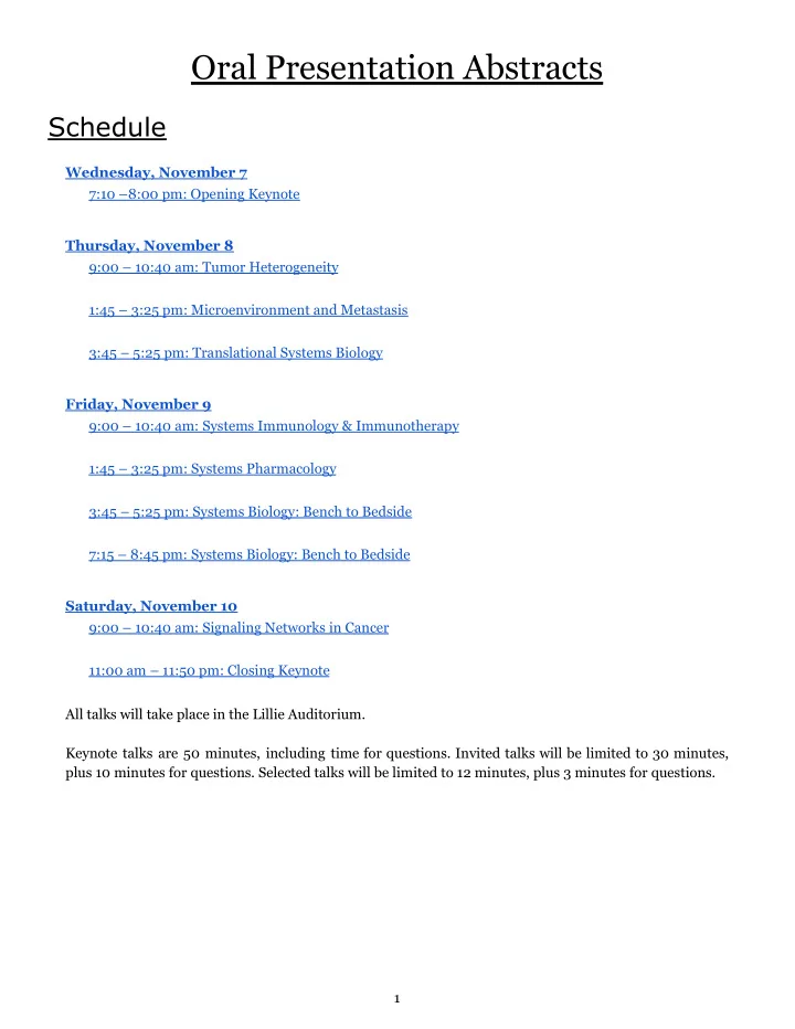

Oral Presentation Abstracts Schedule Wednesday, November 7 7:10 –8:00 pm: Opening Keynote Thursday, November 8 9:00 – 10:40 am: Tumor Heterogeneity 1:45 – 3:25 pm: Microenvironment and Metastasis 3:45 – 5:25 pm: Translational Systems Biology Friday, November 9 9:00 – 10:40 am: Systems Immunology & Immunotherapy 1:45 – 3:25 pm: Systems Pharmacology 3:45 – 5:25 pm: Systems Biology: Bench to Bedside 7:15 – 8:45 pm: Systems Biology: Bench to Bedside Saturday, November 10 9:00 – 10:40 am: Signaling Networks in Cancer 11:00 am – 11:50 pm: Closing Keynote All talks will take place in the Lillie Auditorium. Keynote talks are 50 minutes, including time for questions. Invited talks will be limited to 30 minutes, plus 10 minutes for questions. Selected talks will be limited to 12 minutes, plus 3 minutes for questions. 1
Wednesday, November 7 7:10 –8:00 pm: Opening Keynote Christina Leslie (Memorial Sloan Kettering Cancer Center) Thursday, November 8 9:00 – 10:40 am: Tumor Heterogeneity Chaired by Tenley Archer (Boston Children’s Hospital) Shannon Mumenthaler (University of Southern California) Unlikely suspects: deciphering the functional heterogeneity of fibroblasts in cancer Cancer is a complex adaptive system orchestrated by the interactions between tumor cells and their microenvironment. In particular, cancer-associated fibroblasts (CAFs), the dominant cellular component of the tumor stroma, are often associated with a poor prognosis and play an important role in tumor progression for a number of cancers. While significant literature has highlighted the influence of CAFs on cancer cell phenotypes including tumor cell proliferation and invasion, the role of CAF heterogeneity on treatment response remains largely understudied. Additionally, preclinical treatment studies often focus on drug-induced changes to tumor cells with little investigation into the impact on surrounding stromal cells. To advance our biological understanding of cancer and improve treatment efficacy, we are utilizing quantitative high-content imaging coupled with more physiologically-relevant patient-derived model systems to illuminate the dynamic interactions between cancer cells and their microenvironment. These studies are aimed at increasing our understanding of the functional and therapeutic utility of CAFs by leveraging expertise across disciplines. Our lab has developed several imaging-based workflows, combined with machine learning and other image analysis techniques, to rapidly and accurately classify cell types and cell behaviors within heterocellular populations. Using these approaches, we have identified cancer-associated fibroblasts as a source of environment mediated drug resistance in colorectal cancer. Specifically we discovered a novel mechanism by which drug treated CAFs render adjacent tumor cells resistant to anti-EGFR therapy. Paul Macklin (Indiana University) Open source software for studying 3D multicellular cancer systems biology in high throughput Key cancer processes – such as microenvironmental- and therapy-driven cell adaptations, invasion, and metastasis – take place not just within single cancer cells, but within multicellular systems. In these systems, multiple cell types live and communicate in complex biochemical and biomechanical environments. Therapies perturb these highly nonlinear systems, sometimes with unexpected results including side effects and treatment failure. To improve treatment success, we need to study not just 2
cancer cells in isolation, but also understand and control the multicellular system. Computational models can act as 'virtual laboratories,' where we can systematically study the multicellular systems biology of cancer. The ideal such laboratory would include cell and tissue biomechanics, biotransport of multiple chemical substrates including signaling factors, and many interacting cells. We recently developed and released PhysiCell ( http://dx.doi.org/10.1371/journal.pcbi.1005991 ), an open source platform for 3-D multicellular systems biology. With this platform, desktop workstations can routinely simulate systems of ten or more cell-secreted chemical signals and tissue substrates, along with 10^5 to 10^6 cells that grow, divide, die, secrete chemical signals, move, exchange mechanical forces, and remodel their tissue microenvironment. We demonstrate PhysiCell in 2D and 3D simulation examples that examine (1) how mechanical interactions between cancer cells and the liver parenchyma can affect the successful seeding of colon cancer metastases, (2) how biochemical and biomechanical interactions between motile and non-motile breast cancer cells can impact tissue invasion, (3) potential designs for synthetic multicellular systems that transport cancer therapeutics, and (4) the critical role of stochastic migration in immune responses to tumors. We will briefly discuss how open source has accelerated PhysiCell's development, and we'll close with early results on using supercomputers to accelerate large-scale computational investigations. In the future, we aim to adapt these systems to drive high-throughput multicellular cancer systems biology. Christopher McFarland (Stanford University) Traversing the fitness landscape of lung adenocarcinoma in vivo using tumor barcoding and CRISPR/Cas9-mediated genome editing Christopher D McFarland, Zoë N Rogers, Ian P Winters, Wen-Yang Lin, Dmitri A Petrov, and Monte M Winslow The evolution of somatic cells into cancer is a rare event. To interrogate the evolutionary outcomes of early-stage tumors within their native environment, we combined tumor barcoding with lenti-Cre and CRISPR/Cas9-based mouse models of lung adenocarcinoma 1 . This technology allows us to precisely track the size of hundreds of tumors of programmable genotype within a single mouse. Strikingly, tumors initiated with the same genetic drivers, at the same time, within the same mouse vary in size by >1,000-fold after only ten weeks of tumor growth. Furthermore, different cancer genotypes exhibit categorically-different tumor size distributions. We propose two simple Markov models of tumor evolution to explain these different size distributions, whereby heavy-tailed tumor size distributions arise when a second transformative event is necessary for advanced tumor size. We then tested these two models by tracking the growth of hundreds of tumor growth trajectories over time using our quantitative barcoding approach in twelve different cancer genotypes. Our findings and model indicate that the likelihood of cancer depends on the variability in growth imparted by driver events, more than their mean effect, as malignancy represent an exceedingly-rare, exceptionally-advanced evolutionary state. 1 Rogers, McFarland, Winters et al (2017). A quantitative and multiplexed approach to uncover the fitness landscape of tumor suppression in vivo. Nature Methods, 14:737-42. B. Bishal Paudel (Vanderbilt University) Drug response epigenetic landscape of BRAF-mutated melanoma cells B. Bishal Paudel, Leonard A. Harris, Keisha N. Hardeman, Arwa A. Abugable, Corey E. Hayford, Darren R. Tyson, Christian T. Meyer, Joshua P. Fessel, and Vito Quaranta 3
Recommend
More recommend