Journal of Sport and Kinetic Movement Vol. I, No. 31/2018 SECONDARY - - PDF document

journal of sport and kinetic movement vol i no 31 2018
SMART_READER_LITE
LIVE PREVIEW

Journal of Sport and Kinetic Movement Vol. I, No. 31/2018 SECONDARY - - PDF document

Journal of Sport and Kinetic Movement Vol. I, No. 31/2018 SECONDARY AND TERTIARY PROFILAXY OF THE MOYAMOYA DISEASE IN CHILDHOOD CASE PRESENTATION Elena BUHOCIU Spiru Haret University, Bucharest Romania Faculty of Physical Education, Sport and


slide-1
SLIDE 1

Journal of Sport and Kinetic Movement Vol. I, No. 31/2018

36

SECONDARY AND TERTIARY PROFILAXY OF THE MOYAMOYA DISEASE IN CHILDHOOD CASE PRESENTATION

Elena BUHOCIU

Spiru Haret University, Bucharest Romania Faculty of Physical Education, Sport and Physical Therapy, Bucharest, Romania *Correspondence address: ebuhociu@yahoo.com Abstract: The Moyamoya syndrome (also known as the Moyamoya disease) consists of a series of malformations and

congenital modifications of the cerebral arteries and occlusions and narrowing of the terminal carotid artery bifurcation. The term Moyamoya comes from the Japanese language and means "puff smoke". This describes the arteriography of cerebral artery changes, which emit numerous small-diameter collateral vessels with fragile walls. The diagnosis of Moyamoya disease is established after excluding other genetic or congenital causes of such vascular malformations. These vessels with increased fragility often have parietal lesions at increased risk of rupture and secondary stroke. In the present paper we aim to present the case of a child diagnosed with the Moyamoya disease who suffered two vascular accidents and is currently in the recovery process. The treatment is complex and consists of surgery and rehabilitation. Depending on the location of the cerebral malformations, clinical manifestations are diverse. In the present case, they are motor, but the child also has speech disorders, even if the intelligence is preserved. Keywords: Vascular malformations, child stroke, rehabilitation.

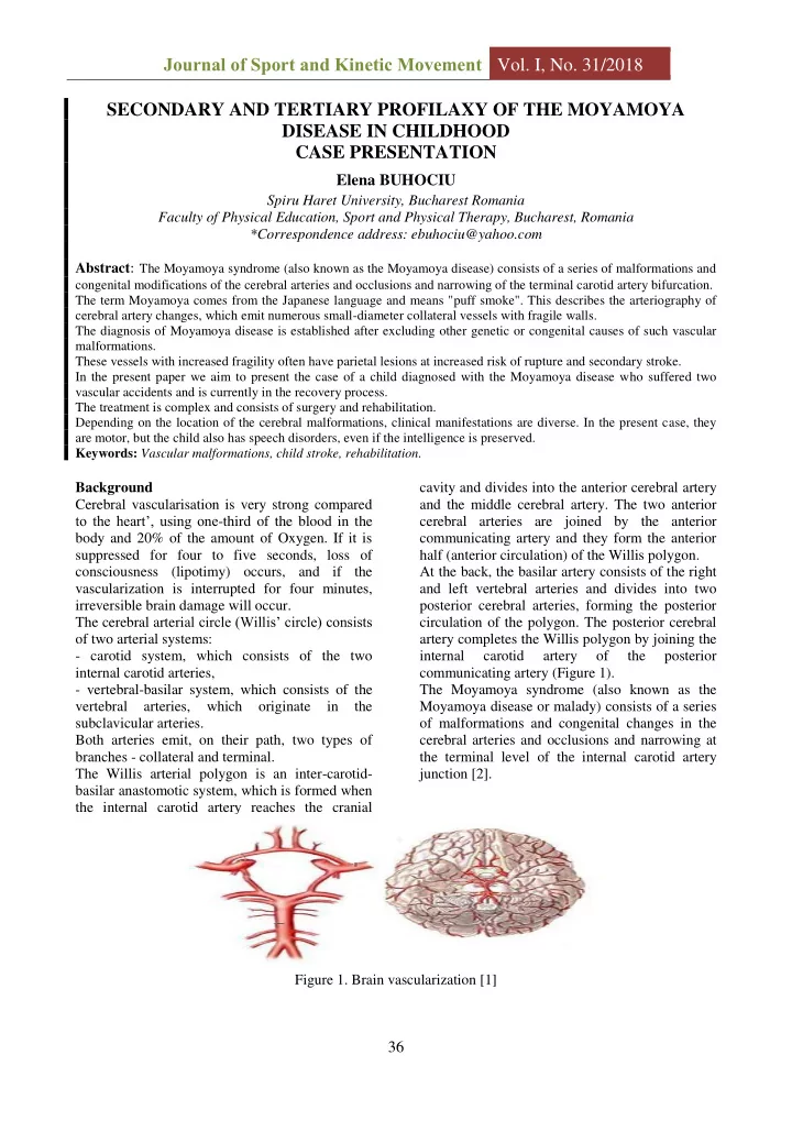
Background Cerebral vascularisation is very strong compared to the heart’, using one-third of the blood in the body and 20% of the amount of Oxygen. If it is suppressed for four to five seconds, loss of consciousness (lipotimy) occurs, and if the vascularization is interrupted for four minutes, irreversible brain damage will occur. The cerebral arterial circle (Willis’ circle) consists

  • f two arterial systems:
  • carotid system, which consists of the two

internal carotid arteries,

  • vertebral-basilar system, which consists of the

vertebral arteries, which

  • riginate

in the subclavicular arteries. Both arteries emit, on their path, two types of branches - collateral and terminal. The Willis arterial polygon is an inter-carotid- basilar anastomotic system, which is formed when the internal carotid artery reaches the cranial cavity and divides into the anterior cerebral artery and the middle cerebral artery. The two anterior cerebral arteries are joined by the anterior communicating artery and they form the anterior half (anterior circulation) of the Willis polygon. At the back, the basilar artery consists of the right and left vertebral arteries and divides into two posterior cerebral arteries, forming the posterior circulation of the polygon. The posterior cerebral artery completes the Willis polygon by joining the internal carotid artery

  • f

the posterior communicating artery (Figure 1). The Moyamoya syndrome (also known as the Moyamoya disease or malady) consists of a series

  • f malformations and congenital changes in the

cerebral arteries and occlusions and narrowing at the terminal level of the internal carotid artery junction [2]. Figure 1. Brain vascularization [1]

slide-2
SLIDE 2

Journal of Sport and Kinetic Movement Vol. I, No. 31/2018

37

Occlusions are mostly bilateral and are localized

  • n the arteries that form the Willis arterial

polygon [3] at the base of the brain, resulting in the development of a neo-vascular arteriole and capillary network with increased fragility. These vessels with increased fragility often have parietal lesions at increased risk of rupture and secondary

  • stroke. The diagnosis of Moyamoya disease is

established after excluding other genetic or congenital causes of such vascular malformations. The term Moyamoya comes from the Japanese language and means "smoke cloud". This describes the arteriography of cerebral artery changes, which emit numerous small-diameter collateral vessels with fragile walls. According to

  • prof. Burunsus (State University of Medicine and

Pharmacy “Nicolae Testemiţanu” - Chişinău), the term would refer to "a thin fog-like appearance, a fine, fluffy image that is given by the basal anastomotic vascular network" [4] (Figure 2). Figure 2. Angiographic image - Moyamoya syndrome [5, 6] The disease is also known as the juvenile

  • cclusive disease of the Willis polygon, cerebral

brain jungle brain telangiectasia, or chronic cerebral occlusive vasculopathy [5], but in our

  • pinion

none

  • f them

fully express the etiopathogenic aspects and manifestations of the

  • disease. On the other hand, the condition is not

within the group of cerebral vasculitis [6]. The reported incidence of the disease in the USA is of 0.086 per 100,000 people [7]. It was first described in 1957 in Japan as hypoplasia of internal carotid arteries. In this country, the incidence is about 4 cases per 100,000 people. Both European countries, as well as other countries of the Far East and the American continent, have been reported. An incidence of 0.35 per 100,000 people is reported in Europe. The disorder may be unilateral or bilateral and may occur at any age [8]. Anatomopatologically, it is described as the tunica indima’s thickening in the terminal parts of the internal carotid vessels at bilateral level. Proliferation of the intima may also contain lipid

  • deposits. The anterior, middle and posterior

arteries that form the Willis polygon may exhibit varying degrees of stenosis or occlusion, this being associated with thickening of the tunica intima, thinning of the tunica media and the fragmentation of the elastic membrane. Once the artery occlusion begins, it is progressive, in some people it can cause repeated vascular cerebral attacks that lead to severe functional sequelae, or even death, and in some there may be no

  • symptoms. The Moyamoya disease initially

causes constrictions in the internal carotid artery, but often the anterior and middle artery can be affected. The Moyamoya-specific arterial constriction is different from the one caused by arteriosclerosis. In arteriosclerosis, the walls of the arteries are damaged, leading to cell deposition, while in Moyamoya, the internal layer of the carotid artery proliferates inside the arterial lumen. Within the artery, blood clots are also formed. The abnormal vascular network develops especially in the carotid apexes, consisting of small and thin vessels along the affected area. This network, being very fragile, is exposed to the risk of arterial rupture (Figure 3).

slide-3
SLIDE 3

Journal of Sport and Kinetic Movement Vol. I, No. 31/2018

38

Figure 3. Vascular Neoformation Network - Moyamoya Syndrome [9] The etiology of the disease is less well known - it seems to be related to a number of modified genes found on chromosomes 3, 6, 8, 17, 23. In 10% of cases, the hereditary factor is established. This is also enforced by the presence of HLA-type antigens in the blood of patients with the Moyamoya syndrome. Prevalence: The disease is more common in women than in males. It can also be observed that infantile forms are more frequent than in adults. The Symptomatology is unspecific, of neurologic and vascular types: persistent migraine headache, loss of consciousness, nonspecific changes in vision (phosphenes, scotomas, temporary loss of vision), hemiparesis, speech disorders, personality changes, seizures (especially in children), involuntary fine movements of extremities etc. Although initially the child's intelligence may be normal, a slow and progressive mental deterioration may occur over time. These symptoms may appear gradually, diminishing over time, or may have a sudden

  • nset, but are persistent.

Evolution is progressive and occurs in several stages, which are based

  • n

angiographic examinations:

  • Stage I: narrowing of the internal carotid artery’s

bifurcation;

  • Stage II: dilatation of the anterior and middle

cerebral arteries;

  • Stage III: narrowing of the internal carotid

artery’s bifurcation, of the anterior cerebral artery,

  • f the middle artery and the appearance of the

basal collateral circulation - it’s the stage in which the condition is usually diagnosed;

  • Stage IV: internal carotid artery occlusion and

development of collateral circulation;

  • Stage V: accentuation of internal carotid artery
  • cclusion and diminution of blood flow through

unformed collateral vessels;

  • Stage VI: disappearance of specific collateral

circulation and complete occlusion of internal carotid arteries. The major complication of the disease is stroke, which may be single or multiple. It is due, on the

  • ne hand, to the degradation of vascular walls,

leading to vasculitis, favoring the formation of clots and ischemia. On the other hand, the fragility

  • f newly formed vessels causes their breakage and

the production of haemorrhagic strokes. Pediatric cerebral accident may occur at two different time intervals. The first is the perinatal interval, which includes both fetal (prenatal) and neonatal - from birth to 28 days. The greatest risk is in the first year of life. The second interval is childhood, which starts on day 29 and continues until the age of 18. Vascular ischemic stroke is the most common in this category of patients. Diagnosis of certainty requires the patient’s family and medical history and careful examination of symptoms, along with detailed laboratory and imaging (MRI, angiography) investigations. Early diagnosis is, unfortunately, almost impossible because angiography presents some risk and can’t be included in the screening examinations. The differential diagnosis of the pediatric Moyamoya syndrome is achieved with other conditions, during which cerebral vascular accidents can occur: siclemia, other blood diseases, congenital heart disease, infections (meningitis), cerebral trauma, dehydration etc. Drug treatment is palliative and only produces an improvement in symptomatology. Surgical treatments are the most effective, because they reduce the likelihood of strokes. The procedures are complex and consist of arterial, muscular or epiplotic autotransplants, the vessels developed at this level providing an additional cerebral blood flow [10]. Hypotheses Physical therapy can’t be used as a curative treatment for Moyamoya disease, but it is indispensable for both for recovery of spastic hemiplegia that installed after repeated strokes and after revascularization surgery.

slide-4
SLIDE 4

Journal of Sport and Kinetic Movement Vol. I, No. 31/2018

39

Materials and Method The patient studied in this work is 5 years old. The child’s symptomatology of the child began in 2016, through three paroxysmal manifestations with bilateral elementary motor deficit in the lower limbs. Anticoagulant treatment has been instituted to prevent ischemic recurrences. In May 2017, following an MRI and cerebral angiography, distal stenosis was identified at the level of both internal carotid arteries, specific for the Moyamoya disease, as well as multiple ischemic degenerative lesions. At the time of admission, the child was conscious, cooperative, temporo-spatial oriented, and the neurological examination offered normal results. No hereditary factor could be identified in the present case, both the parents and the child's brother missing any of the specific clinical and paraclinical symptomatology. Surgical intervention was performed in a mixed team with neurosurgery and vascular surgery

  • specialists. Revascularization was performed by

the procedure called encephalo-myo-synangiosis (EMS), in which small portions of the temporal muscle were attached to the exposed temporal cortex. The patient's evolution after surgery was favorable: it was afebrile and the neurological examination did not change. During the hospitalization, they presented some paroxystic episodes, which consisted of incoherent discourse and verbal repetitions, which is why they began antiepileptic treatment. On the first postoperative day, a computerized cerebral tomography was performed, which revealed a right temporo-occipital ischemic stroke that did not involve deep structures due to the partial occlusion of some branches in the middle and right posterior arteries. Approximately one month after surgery, another cerebral MRI revealed the presence of an ischemic area in the superficial temporo-parietal territory

  • n the left, with an overactive, gyriform aspect,

predominantly at the gray matter’s level. Stenosis in the carotid and cerebral arteries was observed, as well as the presence of multiple collateral circulation patterns specific to the Moyamoya

  • disease. At the admission, the child was agitated

psychomotor-wise, aphasic, showed food intolerance and upper right paresis. Surgery was performed for the second time in a mixed team, neurosurgery and vascular surgery, and revascularization was performed using the same procedure that was previously used, encephalo-myo-sinangiosis. The patient’s evolution after the intervention was favorable, the neurological condition improving. The child still presents hemiparesis, predominantly brachial, was aphasic and afebrile. Two weeks after the surgery, the child was released and transferred to a specialized center for monitoring and for the neuromotor recovery program. Following the neuromotor evaluation, he was diagnosed with spastic right hemiparesis, with a higher deficit in the upper limb - fingers in semiflexion in interphalangeal joints II-III, adducted thumb, without bidigital and tridigital

  • grip. At the level of the inferior limb, hypertonia
  • f sural triceps, level I-II on the Ashworth scale,

with independent walking, was installed; the child can walk without plantar run. They are able to walk on the heel. The patient presented in the paper is at an average stage of spasticity, at which point the spontaneous recovery process ceased. The characteristics of the stage are:

  • increased muscle tone,
  • hypertonia at the level of sural triceps of the

right limb, score 2 on the Ashworth scale,

  • walking is independent, without plantar run,

without running and it is possible to walk on the heel.

  • At upper member level, the second and third

phalanx of the fingers are in semiflexion with adducted thumb,

  • missing bidigital and tridigital grip,
  • motion initiation is improved, but its control is

insufficient due to spasticity. Prior to developing the kinetic program tailored to the needs of the child, it was necessary to set goals to be pursued during recovery. The objectives of kinetotherapy were:

  • inhibition of the abnormal posture and

movement patterns;

  • reducing spasticity;
  • preventing deformities and nefarious attitudes

caused by increased muscle tone;

  • toning of paravertebral muscles and scapular

humerus;

  • restoring

active mobility, strength and coordination, especially at the higher member’s level;

  • recovery of bidigital and tridigital grip at the

right hand,

  • reeducation of walking,
  • balance recovery;
  • gaining

higher degree

  • f

functional independence in self-care.

slide-5
SLIDE 5

Journal of Sport and Kinetic Movement Vol. I, No. 31/2018

40

Parallel to kinetotherapy, which was practiced daily, there were logopedic and occupational therapy programs with specialized staff. The kinetic program consisted of:

  • initial passive mobilizations;
  • the Kabat method;
  • neuro-proprioceptive facilitation techniques;
  • programs of walking and gripping re-education

especially on the right side;

  • muscle toning exercises;
  • coordination exercises etc.

Results and discussions During the kinetic program, the child was very cooperative, although it was necessary to keep to their pace. Many movements were limited, but the patient employed the unaffected upper limb to complete the exercises. After completing the kinetic program applied for this case study, the child used his affected hand much more during the activities than at the beginning, the shoulder extension was greatly improved and even spontaneously performed in

  • ccupational therapy activities.

Spasticity from the upper limb and especially from the hand was reduced in each session after the passive mobilizations. This has made it easier to execute the bidigidal, tridigital, and forceful

  • grip. Initially, the child employed their unaffected

hand to help grippind, afterwards without, however, the maximum amplitude was not

  • reached. Through work-based exercise, adapted

through play, it was possible to extend the fingers. Also, spasticity from sural triceps was reduced by neuro-proprioceptive facilitation techniques, followed by active walking exercises and toning

  • f the lower limb flexing muscles. The plantar run

was closer to physiological and the balance was improved. Conclusions Although very rare in Romania, Moyamoya syndrome poses great problems in establishing certainty in diagnosis and achieving the differential diagnosis. Carriers of this disease are prone to repeated ischemic and / or haemorrhagic stroke. The treatment of choice is the revascularization of the ischemic brain area by surgical intervention. Once the brain injury is installed, the kinetotherapeutic behavior is very similar to that followed in the medical and kinetic recovery of adults. During the recovery sessions, the child became more motivated and more involved in activities. The duration of the sessions has increased gradually since the child was more interested and more focused on recovery exercises. The benefits of kinetotherapy in this case were both motor as well as cognitive and emotional, so the hypothesis formulated at the beginning of the study that rehabilitation treatment for children with spastic hemiplegia is essential for the long- term evolution of subjects has been fully confirmed. References

[1]. http://sanatate.bzi.ro/simptomele-bolii-moya- moya-7309/accessed at 21.09.2018 [2]. Popa, S. (2013). Vascularizația arterială și drenajul venos al creierului. https://anatomie.romedic.ro/ vascularizatia-arteriala-si-drenajul-venos-al-creierului/ accessed at 17.10.2018; [3]. Popa, C. (2016). Boala și sindromul Moyamoya. In http://www.viata-medicala.ro/*article ID_11487- dArt.html/accessed at 16.10.2018; [4]. Burunsus, V., Glavan, I. (2010). Boala Moyamoya: epidemiologie, etiopatogenie, fiziopatologie şi

  • anatomopatologie. In Revista Literaturii. Chișinău. pp.

61-66; [5]. https://www.researchgate.net/figure/Moyamoya- disease-in-a-25-year-old-man-DSA-image-lateral- view-shows-extensive_fig9_ 263899333 /accessed in 12.10.2018; [6].Razek, A.A., Alvarez, H., Bagg, S., Refaat, S., Castillo, M. (2014). Imaging Spectrum of CNS

  • Vasculitis. Publmed. Radiographics. 2014 Jul-Aug;

34(4):873-94. doi: 0.1148/rg.344135028. In https://www.ncbi.nlm.nih.gov/pubmed /25019429/accessed at 10.10.2018; [7].Scott, R.M., Smith, E.R. (2009). Moyamoya Disease and Moyamoya Syndrome. N Engl J Med 2009; 360:1226-1237. DOI: 0.1056/NEJ Mra0804622. In https://www.nejm.org/doi/ full/ 10.1056/nejmra 0804622/accessed in 17.10.2018. [8]. https://www.romaniatv.net/premiera-la- spitalul- bagdasar - un - barbat – diagnosticat - cu- boala-moya-moya-a-fost-operat-cu- succes_149959.html /accessed 13.10.2018. [9]. http://neuroangio.org/neuroangio-topics/ intracranial-collateral-pathways/ accessed at 16.10.2018; [10].https://www.columbianeurosurgery.org/conditions Moyamoya-syndrome/accessed at 17.10.2018.