
Control of CD1d-restricted antigen presentation and inflammation by sphingomyelin Espen Melum 1,2 *, Xiaojun Jiang 2 , Kristi D. Baker 1,3 , M. Fatima Macedo 4,5,6 , Jürgen Fritsch 7,8 , C. Marie Dowds 9 , Jing Wang 10 , Anne Pharo 2 , Arthur Kaser 11 , Corey Tan 1,2 , Catia S. Pereira 4,5 , Samuel L. Kelly 12 , Jingjing Duan 12,17 , Tom H. Karlsen 2 , Mark A. Exley 1 , Stefan Schütze 7 , Dirk M. Zajonc 10,13 , Alfred H. Merrill 12 , Edward H. Schuchman 14 , Sebastian Zeissig 9,15,16,18 and Richard S. Blumberg 1,18 * Invariant natural killer T (iNKT) cells recognize activating self and microbial lipids presented by CD1d. CD1d can also bind non- activating lipids, such as sphingomyelin. We hypothesized that these serve as endogenous regulators and investigated humans and mice deficient in acid sphingomyelinase (ASM), an enzyme that degrades sphingomyelin. We show that ASM absence in mice leads to diminished CD1d-restricted antigen presentation and iNKT cell selection in the thymus, resulting in decreased iNKT cell levels and resistance to iNKT cell-mediated inflammatory conditions. Defective antigen presentation and decreased iNKT cells are also observed in ASM-deficient humans with Niemann – Pick disease, and ASM activity in healthy humans cor- relates with iNKT cell phenotype. Pharmacological ASM administration facilitates antigen presentation and restores the levels of iNKT cells in ASM-deficient mice. Together, these results demonstrate that control of non-agonistic CD1d-associated lipids is critical for iNKT cell development and function in vivo and represents a tight link between cellular sphingolipid metabolism and immunity. I Invariant natural killer T (iNKT) cells are an important lympho- cyte population that sense self- and microbial lipids non-stimulatory CD1d-associated lipid in vitro 9 , leading us to hypo- presented by the major histocompatibility complex (MHC) class thesize that it may regulate CD1d access to potentially agonistic lipids. I-like gly- coprotein CD1d 1 . In response to these antigens, Sphingomyelin is degraded by sphingomyelinases into ceramide iNKT cells rapidly release large arrays of mediators, making and phosphorylcholine 10 . In lysosomes, one of the sites where the them powerful and early modulators of immune pathways 2 . The exchange and loading of lipids onto CD1d takes place 11 , ASM is the self-reactivity of iNKT cells is also critical for their primary enzyme responsible for sphingomyelin degradation 12,13 . In development in the thymus 3 , where iNKT cells are positively light of the non-stimulatory nature of sphingomyelin in vitro 9 , we selected by CD1d-bearing thymocytes 4 . While there have been sought to understand the consequences of sphingomyelin accumu- great efforts to identify CD1d-binding, iNKT cell- activating lation on iNKT cell function. To do so, we used mice with homozy- lipids (that is, lipid antigens 5 ), iNKT cell activation is also gous deficiency in the gene encoding ASM ( Smpd1 – / – ; called ‘ Asm − / − ’ amenable to negative regulation by CD1d-associated lipids that here) 14 that develop age-dependent accumulation of sphingomyelin do not stimulate the iNKT cell antigen receptor (TCR). As and a clinical phenotype resembling ASM deficiency in humans, such, iNKT cell activation is anticipated to be influenced by the known as Niemann – Pick disease (NPD) types A and B, where balance of CD1d-associated antigenic and non-antigenic NPD-A is the more severe form of disease first manifesting during lipids. However, little is known about the functional infancy. We demonstrate that control of the cellular abundance of relevance of non-antigenic lipids that potentially impede sphingomyelin by ASM regulates the presentation of endogenous CD1d-restricted iNKT cell activation. Sphingolipids, which are and exogenous lipid antigens by CD1d in thymocytes and dendritic abundantly present in the cell membrane 6 , are a major class of cells (DCs) and thus the abundance and activation of iNKT cells. CD1d-associated lipids 7,8 . Sphingomyelin, a dominant In the absence of ASM in mice, sphingomyelin levels increased in sphingolipid in mammals, has been reported to be a 1 Gastroenterology Division, Department of Medicine, Brigham and Women’s Hospital, Harvard Medical School, Boston, MA, USA. 2 Norwegian PSC Research Center, Department of Transplantation Medicine, Division of Surgery, Inflammatory Diseases and Transplantation, Oslo University Hospital, Rikshospitalet, and University of Oslo, Oslo, Norway. 3 Department of Oncology, Medical Microbiology and Immunology, University of Alberta, Edmonton, Alberta, Canada. 4 i3S Instituto de Investigação e Inovação em Saúde, Universidade do Porto, Porto, Portugal. 5 Instituto de Biologia Molecular e Celular, Universidade do Porto, Porto, Portugal. 6 Department of Medical Sciences, University of Aveiro, Aveiro, Portugal. 7 Institute of Immunology, Christian- Albrechts University, Kiel, Germany. 8 Department of Infection Prevention and Infectious Diseases, University of Regensburg, Regensburg, Germany. 9 Department of Internal Medicine I, University Medical Center Schleswig-Holstein, Christian-Albrechts University, Kiel, Germany. 10 Division of Immune Regulation, La Jolla Institute for Immunology, La Jolla, CA, USA. 11 Division of Gastroenterology and Hepatology, Department of Medicine, University of Cambridge, Addenbrooke’s Hospital, Cambridge, UK. 12 School of Biological Sciences and the Petit Institute for Bioengineering and Biosciences, Georgia Institute of Technology, Atlanta, GA, USA. 13 Department of Internal Medicine, Faculty of Medicine and Health Sciences, Ghent University, Ghent, Belgium. 14 Department of Genetics & Genomic Sciences, Icahn School of Medicine at Mount Sinai, New York, NY, USA. 15 Department of Medicine I, University Medical Center Dresden, Technische Universität Dresden, Dresden, Germany. 16 Center for Regenerative Therapies Dresden, Technische Universität Dresden, Dresden, Germany. 17 Present address: Human Aging Research Institute, School of Life Sciences, Nanchang University, Nanchang, China. 18 These authors jointly supervised this work: Sebastian Zeissig, Richard S. Blumberg. *e-mail: espen.melum@medisin.uio.no; rblumberg@bwh.harvard.edu
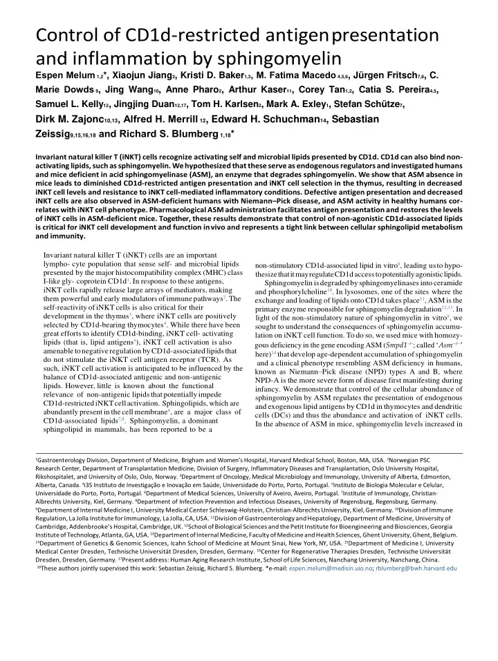
a b 10 5 10 5 Asm – / – spleen WT spleen ASM activity in WT mice ( n = 4) 10 4 10 4 Spleen 1.2% 0.085% Thymus PBS57-CD1d 10 3 10 3 Colon tetramer-PE Liver DCs 10 2 10 2 0 0 10 20 30 40 50 ASM activity (nmol h – 1 mg – 1 ) 0 10 2 10 3 10 4 10 5 0 10 2 10 3 10 4 10 5 CD3-APC c d Thymus Spleen Liver Thymus Spleen Liver ** iNKT cells (%) iNKT cells (%) ** 2.5 × 10 5 4 × 10 5 4 × 10 5 2.0 80 0.5 No. iNKT cells No. iNKT cells No. iNKT cells 2.0 × 10 5 iNKT cells (%) 3 × 10 5 3 × 10 5 0.4 1.5 60 1.5 × 10 5 0.3 2 × 10 5 2 × 10 5 1.0 40 1.0 × 10 5 0.2 1 × 10 5 1 × 10 5 0.5 20 5.0 × 10 4 0.1 0.0 0 0 0.0 0.0 0 e 10 5 Asm – / – spleen 10 5 WT spleen 10 4 10 4 0.39% 0.046% PBS57-CD1d 10 3 tetramer-PE 10 3 10 2 10 2 0 0 10 2 10 3 10 4 10 5 0 10 2 10 3 10 4 10 5 CD3-APC Fig. 1 | Acid sphingomyelinase-deficient mice have a reduced number of iNKT cells. a , ASM activity was measured in tissues from wild-type (WT) mice ( n = 4) using a colorimetric assay in tissue lysates generated by repeated freeze – thaw cycles from the indicated tissues. The results are representative of two independent experiments. b , Representative flow cytometry of lymphocytes from spleens in WT and Asm − / − mice visualizing the number of iNKT cells as defined by a PBS57-loaded CD1d tetramer and CD3. c , Percentages of iNKT cells among lymphocytes in the thymus, spleen and liver of age- and sex-matched WT ( n = 5) and Asm − / − ( n = 4) mice defined by a PBS57-loaded CD1d tetramer and CD3. The results are representative of three independent experiments. d , Absolute numbers of iNKT cells in the thymus, spleen and liver of age- and sex-matched WT ( n = 5) and Asm − / − ( n = 5) mice defined by a PBS57-loaded CD1d tetramer and TCR β . The results are representative of three independent experiments. e , Representative flow cytometry of lymphocytes from spleens in WT and Asm − / − mice at 2 weeks of age visualizing the number of iNKT cells as defined by a PBS57-loaded CD1d tetramer and CD3. In all panels, the mean values are shown with the error bars representing the s.e.m. P values were calculated by a two-sided Student’s t -test. * P < 0.001, ** P < 0.0001. periphery 16 . Consistent with this being functionally impor- tant, flow hematopoietic cells, resulting in decreased CD1d-restricted antigen cytometry (gating strategies in Supplementary Fig. 1) presentation, impaired iNKT cell development in the thymus and reduced abundance and activation of iNKT cells, defects that were reversed by the transfer of wild-type bone marrow or administra- tion of recombinant human ASM (rhASM). These observations can be extended to humans with or without NPD and establish ASM, through its control of sphingomyelin levels, as an important regula- tor of iNKT cells with potential therapeutic implications. Results ASM is active in the hematopoietic system and required for iNKT cell development. Although it is well established that ASM is expressed in tissues where the clinical phenotypes of NPD-A and NPD-B are most prominent, such as the liver and brain as well as in some hematopoietic cells, such as macrophages 15 , little is known about its function in the immune system. Therefore, we first inves- tigated ASM activity and found it to be demonstrable in a vari- ety of parenchymal (colon and liver) and hematopoietic (spleen, thymus and dendritic) cells, with the highest levels in DCs (Fig. 1a), a critical CD1d-expressing antigen-presenting cell (APC) in the
Recommend
More recommend