
Curr Pediatr Res 2012; 16 (1): 15-18 Ataxia telangiectasia: Serological Presentation Chani Gupta, Reshu Tewari, S.M. Natu, Pushpa Tondon, Raj Mehrotra Department of Pathology, C S M Medical University (Earlier King George’s Medical University), Lucknow, U.P., India Abstract Ataxia telangiectasia is a rare autosomal recessive multisystem disorder,having an incidence of 1:40,000 to 1:100,000 with an equal ratio in males and females, characterized by cerebel- lar ataxia, variable immunodeficiency, oculocutaneous telangiectasia, increased x ray hyper- sensitivity and susceptibility to malignancies. The causative gene has been localized to chromosome 11q22-23. Here a case of an 8 year old boy is described who presented with progressively increasing gait difficulties, immunological and ocular manifestations, bilateral CSOM, and abdominal tuberculosis. The case an almost classical presentation of ataxia te- langiectasia, highlights the diagnostic work up and the serological findings for early detec- tion and genetic counseling of the parents. Key Words : ataxia telangiectasia, alpha-fetoprotein, immunodeficiency Accepted October 21 2011 Introduction enzyme linked immunosorbent asssay, serum IgE and IgA levels as well as other relevant serological investigations. Ataxia telangitectasia (ATM) is a rare hereditary neu- The results showed elevated AFP levels with no rise in rodegenerative disease usually found in early childhood CEA levels. Patient’s serum also showed slight elevation [1] . It is characterized by a progressive cerebellar in co- in liver function tests, not a well documented findings in ordination, with the patient being wheel chair bound by patients of ataxia telangiectasia. 10-11yrs of age [1]. Oculocutaneous involvement in the form of telangiectasia may manifest itself by 3-6 yrs of Case presentation age. The patient usually suffers from repeated sinopul- monary infections attributed to variable reduction in lev- els of serum and secretory IgA and IgE and are one of the An 8 year old boy presented with progressively increas- major cause of mortality in such children . The causative ing difficulty in walking, and coordinating hand move- ATM gene has been localized to band 11q22-23. ATM ments since two years and also complained of reddening has sequence homology to a family of proteins that are of eyes. He was being treated for frequent cough and cold. related to the phosphatidylinositol-3-OH-kinases (PI (3) The boy had a past history of bilateral chronic suppurative K) [1], and have a role in DNA repair, which is presumed otitis media and taking antitubercular treatment for Koch to be responsible for increased susceptibility to malignan- abdomen. There was no history of delay or abnormality in cies. Serum levels of oncofetoproteins like AFP and CEA developmental milestones. are found to be increased. There was no significant history of a similar disease in The cause of elevated AFP has been attributed to imma- any sibling or any other family member. On examination ture liver, due to a defective interaction between ectoder- sclera of both the eyes revealed dilated blood vessels (Fig. mal 1). Here we present a case of a child who complained of pro- Neuromotor examination showed incoordination of gressive motor dysfunction, immunological dysfunction movement more marked in lower limbs. Deep tendon re- and ocular manifestations of telangiectasias, and was on flexes were normal. medication for frequent cough and cold. We estimated his as well as his mothers’ serum AFP and CEA levels using No abnormality seen on CT scan. Curr Pediatr Res 2012 Volume 16 Issue 1 15
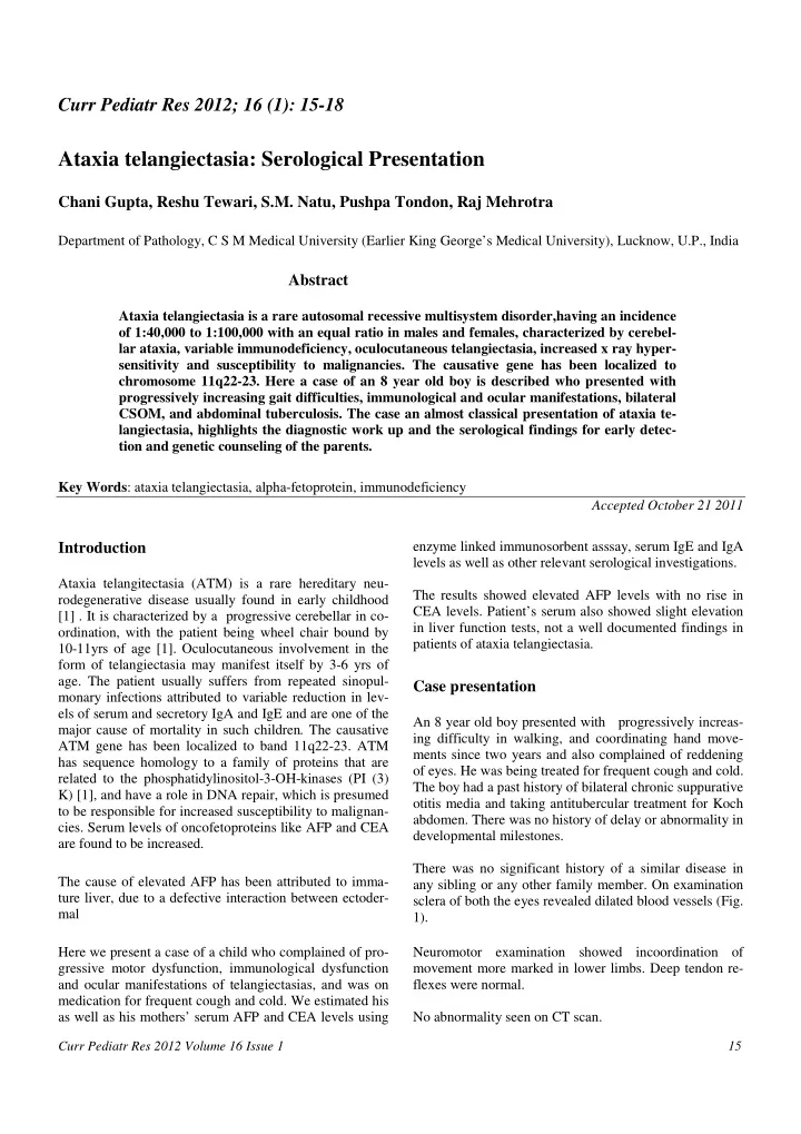
Gupta/ Tewari/ Natu/Tondon/ Mehrotra Figure 1: Shows conjunctival telengiectasias (network of dilated blood vessels) present in bilateral eyes, a characteris- tic finding in Ataxia Telengiectasia Table 1. Details of laboratory investigation. showed Patient Mother Normal Sr. Alpha-fetoprotein 140.6 0.73 <8.5ng/ml Sr. CEA 5.16 4.3 <5ng/ml Sr. IgA 1.610 3.040 0.52-4.68IU/ml Sr. IgE 4.0 259.5 <1.5-378iu/ml Sr. Alkaline Phosphate 358 55 15-112Iu/L SGPT 124 48 9-40U/L SGOT 85 38 9-40IU/L Sr Bilirubin(total) 0.7 0.5 0.5-1.2mg/dl Sr. Albumin 4.1 2.0 3.5-5.0g/dl Sr Protein 6.6 7.1 6.0-8.0g/dl Sr. Urea 20.66 0.5 15-42mg/dl Sr. Creatinine 0.59 0.85 0.5-1.2mg/dl Sr. LDH 299.2 291.9 240-480IU/L ND * Vitamin B12 644.0pg/ml 211-911pg/ml ND * Creatinine Phosphokinase 32.3 25-200 Discussion only for the patient but also for the parents and siblings. Since it is an autosomal recessive disorder, there are 25% Tlthough ataxia telangiectasia has a poor prognosis, with chances of further offsprings being affected .Furthermore no cure and high mortality being usually due to sinopulm- there are increased chances of breast cancer in female monary infections, early diagnosis has implications not 16 Curr Pediatr Res 2012 Volume 16 Issue 1
Recommend
More recommend