
5/10/2013 1
Dynamic Evaluation of Pivot-Shift Kinematics in Physeal-Sparing Pediatric Anterior Cruciate Ligament Reconstruction Techniques
Mark Sena James Chen, MD Ryan Dellamaggioria, MD Dezba G. Coughlin, PhD Jeffrey C. Lotz, PhD Brian T. Feeley, MD Mark Sena PhD candidate Orthopaedic Bioengineering Lab Advisers: Jeffrey Lotz Brian Feeley
May 10, 2013 Orthopaedic Surgery
Am J Sports Med. 2013 Apr; 41(4):826-34
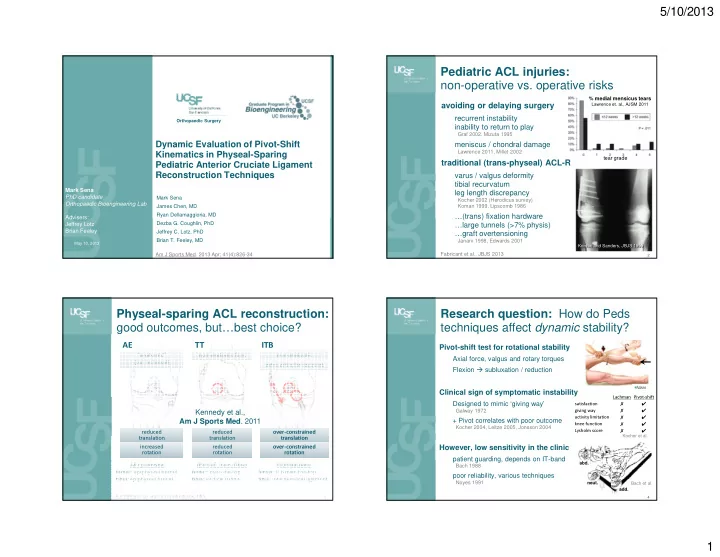
Pediatric ACL injuries: non-operative vs. operative risks
avoiding or delaying surgery
recurrent instability inability to return to play
Graf 2002, Mizuta 1995
meniscus / chondral damage
Lawrence 2011, Millet 2002
traditional (trans-physeal) ACL-R
varus / valgus deformity tibial recurvatum leg length discrepancy
Kocher 2002 (Herodicus survey) Koman 1999, Lipscomb 1986
…(trans) fixation hardware …large tunnels (>7% physis) …graft overtensioning
Janarv 1998, Edwards 2001
2
% medial mensicus tears
Lawrence et. al., AJSM 2011
tear grade
Koman and Sanders, JBJS 1999 Koman and Sanders, JBJS 1999
Fabricant et al., JBJS 2013
Physeal-sparing ACL reconstruction: good outcomes, but…best choice?
3
All Epiphyseal femur: epiphyseal tunnel tibia: epiphyseal tunnel (Partial) Trans-Tibial femur: over-the-top tibia: vertical tunnel Iliotibial Band femur: ITB over-the-top tibia: intermeniscal ligament
Illustrations by Rosanna Wustrack, MD.
“anatomic” (but technical) “anatomic” (but technical) non-anatomic fem. non-anatomic fem. non-anatomic non-anatomic extra-articular restraint extra-articular restraint
Kennedy et al., Am J Sports Med. 2011
reduced translation reduced translation reduced translation reduced translation
- ver-constrained
translation
- ver-constrained
translation increased rotation increased rotation reduced rotation reduced rotation
- ver-constrained
rotation
- ver-constrained
rotation
ITB TT AE
Research question: How do Peds techniques affect dynamic stability?
Pivot-shift test for rotational stability
Axial force, valgus and rotary torques Flexion subluxation / reduction
Clinical sign of symptomatic instability
Designed to mimic ‘giving way’
Galway 1972
+ Pivot correlates with poor outcome
Kocher 2004, Leitze 2005, Jonsson 2004
However, low sensitivity in the clinic
patient guarding, depends on IT-band
Bach 1988
poor reliability, various techniques
Noyes 1991 Lachman Pivot-shift satisfaction ✗ ✔ giving way ✗ ✔ activity limitation ✗ ✔ knee function ✗ ✔ Lysholm score ✗ ✔
Kocher et al. abd. add. neut.
4
Bach et al.