
SLIDE 1
4/19/2013 Dennis Bandyk, MD I have no disclosures Is Carotid - - PowerPoint PPT Presentation
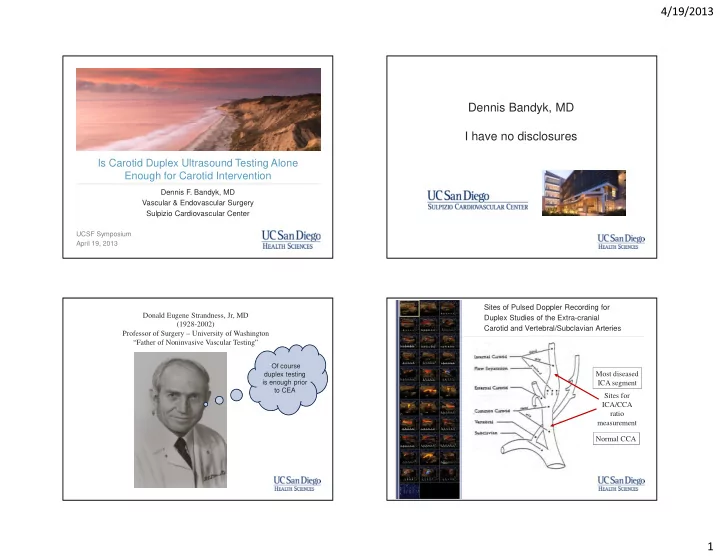
4/19/2013 Dennis Bandyk, MD I have no disclosures Is Carotid Duplex Ultrasound Testing Alone Enough for Carotid Intervention Dennis F. Bandyk, MD Vascular & Endovascular Surgery Sulpizio Cardiovascular Center UCSF Symposium No
Sacco, R. L. N Engl J Med 2001;345:1113-1118
>50% DR >60% DR >70% DR >80% DR
Beach KW, et al Vasc & Endovasc Surg 2012