1
play

1 Major Clinical Sub-types of Scleroderma Major Clinical Sub-types - PDF document

Clinical Features of Scleroderma (Systemic sclerosis; SSc) Scleroderma Chronic multisystemic disease characterized by vasculopathy, variable degree of inflammation, and fibrosis Incidence 3.7-22.8 cases/million Typical facial features in


  1. Clinical Features of Scleroderma (Systemic sclerosis; SSc) Scleroderma • Chronic multisystemic disease characterized by vasculopathy, variable degree of inflammation, and fibrosis • Incidence 3.7-22.8 cases/million Typical facial features in • Female:male 5:1 advanced SSc • Pulmonary fibrosis common, severe in 16%. Pulmonary hypertension occurs in 50% of cases and can lead to cor pulmonale. Pulmonary complications are the leading cause of death in this disease. Digital ulceration from vascular damage CREST: Calcinosis, Raynaud’s phenomenon, Organ Involvement in Scleroderma Sclerodactyly, Esophageal dysmotility, Telangiectasias Telangiectasias Ranaud’s phenomenon Calcinosis Differential organ involvement in SSc. The earliest pathological feature of SSc in the skin is vasculopathy with endothelial cell activation. Later, inflammation develops and finally fibrosis is prominent. Similar processes are likely to occur in all lesional tissues, leading to organ dysfunction. Cardiac, renal, lung and gut complications are the main causes of SSc-related mortality. Sclerodactyly From: Denton and Black Trends Immunol. 26:596, 2005 
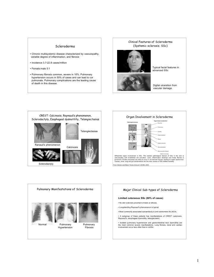
Pulmonary Manifestations of Scleroderma Major Clinical Sub-types of Scleroderma Limited cutaneous SSc (60% of cases) • No skin sclerosis proximal to knees or elbows. • Longstanding Raynaud's phenomenon is typical. • Most commonly associated autoantibody is anti-centromere Ab (ACA). • A subgroup of these patients has manifestations of CREST (calcinosis, Raynaud's, esophageal dysmotility, telangiectasis). • Isolated pulmonary hypertension and gastrointestinal tract dysmotility are Normal Pulmonary Pulmonary the most common severe manifestations. Lung fibrosis, renal and cardiac Hypertension Fibrosis involvement occur less often than in dcSSc. 1

  2. Major Clinical Sub-types of Scleroderma Major Clinical Sub-types of Scleroderma Scleroderma overlap syndromes Diffuse cutaneous SSc (30% of cases) • Their features include those of limited or diffuse cutaneous SSc, together with • The cardinal feature is skin sclerosis proximal to the knees and elbows. those of one or more additional autoimmune rheumatic diseases, such as polyarthritis, myositis or SLE. • Raynaud's phenomenon is universal but might manifest simultaneously or shortly after the development of skin sclerosis. • Often associated with anti U1-ribonucleoprotein (U1-RNP), U3-RNP or polymyositis-scleroderma (PM-Scl) autoantibody reactivity. • Inflammatory features are prominent during the first 3 years of disease. Skin involvement often diminishes within two years. Localized scleroderma • ACA is rarely present, anti-Scl-70 (topoisomease-1) or anti-RNA polymerase is typical. • Morphea scleroderma causes patches of hard skin that can persist for years. • There is a high frequency of interstitial lung disease, renal crisis and cardiac • Linear scleroderma causes bands of hard skin across the face or an involvement. extremity, rarely involving muscle or bone • Typically carries a good prognosis The Role of B Cells in Scleroderma Genetics of Scleroderma • Family history associated with increased risk of developing disease, but risk is only 1% for any individual. • Concordance for both mono- and dizygotic twins is 5%. However, gene expression profiling of cultured dermal fibroblasts from monozygotic twins of an index case show a similar pro-fibrotic “signature” 46% of the time. • Genetic studies indicate an association of scleroderma with polymorphisms in the promoters of the TNF , MCP-1, and CD19 genes. • Although genetic studies have suggested an association of the HLA- DRB1*01 allele with anti-centromere antibodies, association of ACA with In the blood of patients with scleroderm a, naive B cells are increased in number, w hile m emory polymorphisms in the TNF promoter was even stronger, suggesting B cells and plasmablasts/ early plasma cells are diminished. Memory B cells express higher levels linkage disequilibrium. of CD80 and CD86, and thus are chronically activat ed, possibly due t o CD19 over- expression. CD95 ex pr ession is also increased on mem or y B cells, leading to augmented CD95- m ediated apopt osis. The cont inuou s loss of m em ory B cells an d plasmablast s/early plasm a cells m ay increase naive B-cell production in bone marrow to m aintain the B-cell hom eostasis in system ic sclerosis. From: Fujimoto and Sato: Curr. Opin. Rheumatol . 17: 746, 2005. The Role of B Cells in Scleroderma Do B Cells and Autoantibodies Play a Causative Role in Scleroderma? Legend From: Fujimoto and Sato: Curr. Opin. Rheumatol . 17: 746, 2005 2

  3. Autoantibodies to the PDGF Receptor Stimulates Production of Reactive Oxidants Oxidant production triggered by sera-derived IgG upon incubation with fibroblasts over-expressing the PDGF receptor. N, normal controls; SSc, scleroderma; PRP, primary Raynaud’s phenomenon ; SLE, systemic lupus erythematosis; RA, rheumatoid arthritis; ILD, interstitial lung disease; AG 1296, PDGFR kinase inhibitor. From: Baroni et al., New Engl. J. Med. 354:2667, 2006. Auto-antibodies to the PDGF Receptor: Scleroderma-derived Antibodies to the PDGF Receptor The Pathogenesis of Scleroderma Revealed? Stimulate Cell Signaling and Collagen Production IP: PDGF receptor Blot Legend: FCS, fetal calf serum; AG1478, EGFR tyrosine kinase inhibitor; AG1296, PDGFR kinase Unlike normal fibroblasts, fibroblasts in scleroderma increase the expression of PDGFR in respon se to TGF- � , inhibitor rendering the cells more sensitive to PDGF. The Ras–ERK1/2–ROS signaling pathway is triggered by PDGF or anti- PDGFR, which then activates NADPH oxidase (NOX1) to produce reactive oxygen species (ROS). These, in turn, activate extracellular signal-regulated kinases 1 and 2 (ERK1/2), which induce the H- ras gene. This signaling loop is present in normal fibroblasts but is relatively amplified in fibroblasts in patients with scleroderma. From: Tan, New Adapted from: Baroni et al., New Engl. J. Med. 354:2667, 2006 Engl. J. Med. 354:2709, 2006. Therapeutic Approach to the Potential Therapeutics for Scleroderma Treatment of Scleroderma Based on Insight Into its Pathogenesis If chronic over-stimulation of the PDGFR is required for the development and/or progression of scleroderma, then inhibition of PDGFR kinase activity (AG 1296), Ras post-translational processing (FTI 277), or ERK MAP kinase (PD 98059) may prove therapeutic. From: Baroni et al., New Engl. J. Med. 354:2667, 2006. From: Denton and Black Trends Immunol. 26:596, 2005 3

  4. Fibrosis Can Occur in Any Organ and Examples of Fibrosis Can Lead to Irreversible Organ Damage • Various theories have been proposed for the pathogenesis of fibrosing diseases. One view is that fibrosis represents a pathological variant of wound healing. • An alternate view is that fibrosis is due to “unresolved inflammation.” Normal glomerulus Hepatitis C cirrhosis • Another view is that fibrosis results from an “imbalance” in the activities of proteases and anti-proteases. • Regardless of the precise etiology of fibrosis, the pathological deposition of collagen and other components of the extracellular matrix results from persistence of mesenchymal cells assuming a fibroblastic or myofibroblastic phenotype, Glomerulosclerosis Retroperioneal fibrosis producing copious amounts of components of the ECM. Idiopathic Pulmonary Fibrosis (IPF) Three questions worth pondering… Normal 1. What cells and molecules participate in fibrosis? 2. What is the pathogenesis of fibrosis? 3. Can we intervene therapeutically and retard or IPF reverse fibrosis? From: Best et al., Radiology 228:407, 2003; Wittram et al . Radiographics 23:1057, 2003. Fibrosis Results from the Inappropriate Two Engaging Members of the ECM Deposition of Extracellular Matrix Collagen Fibronectin Laminin Fibronectin Laminin Proteoglycan Integrin 4

Download Presentation
Download Policy: The content available on the website is offered to you 'AS IS' for your personal information and use only. It cannot be commercialized, licensed, or distributed on other websites without prior consent from the author. To download a presentation, simply click this link. If you encounter any difficulties during the download process, it's possible that the publisher has removed the file from their server.

Recommend


More recommend