1 2 Thalassemias : Defect in globin biosynthesis E u r o p e a n R - - PDF document

1 2
SMART_READER_LITE
LIVE PREVIEW

1 2 Thalassemias : Defect in globin biosynthesis E u r o p e a n R - - PDF document

Disclosure Company Relationship Type UCSF Continuing Medical Education - Bluebird Bio Site Principal Investigator - Celgene Site Principal Investigator, consultancy, advisory board Thalassemia in the Asian Community : - La Jolla Pharma Site


slide-1
SLIDE 1

1 | [footer text here]

10/11/19 Ashutosh Lal, MD Professor of Clinical Pediatrics UCSF School of Medicine Program Director, Comprehensive Thalassemia Program UCSF Benioff Children’s Hospital Oakland

UCSF Continuing Medical Education

Thalassemia in the Asian Community: Under-recognized & Under-treated

1

Disclosure

§ Company Relationship Type
  • Bluebird Bio
Site Principal Investigator
  • Celgene
Site Principal Investigator, consultancy, advisory board
  • La Jolla Pharma
Site Principal Investigator
  • Protagonist
Site Principal Investigator
  • T
erumo Site Principal Investigator
  • Agios
Co-Investigator
  • Sangamo
Co-Investigator

2

Thalassemias: Defect in globin biosynthesis

Hemoglobin: Tetramer of (Heme + Globin)

Globin Chains Heme Iron Thalassemia: Reduced formation of globin chains Hemoglobinopathy: Abnormal chains, produced at normal rate Globin deficiency Iron deficiency Porphyrin deficiency α β α β

3

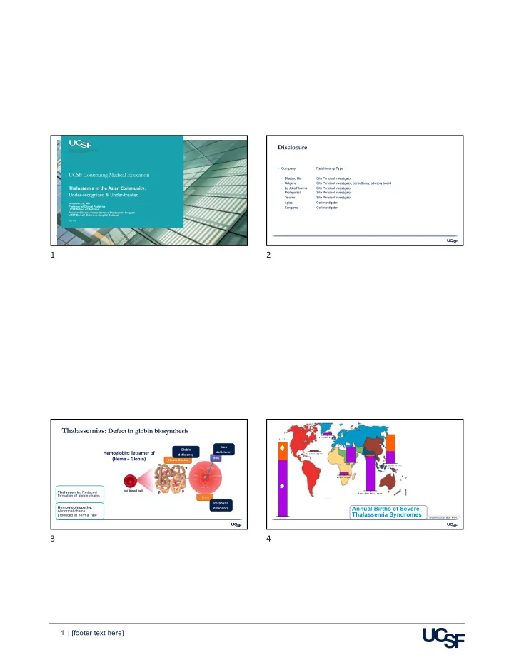
W o r l d 5 5 , 8 7 5 a b A f r i c a n R e g i o n A m e r i c a n R e g i o n E a s t e r n M e d i t e r r a n e a n E u r o p e a n R e g i o n S o u t h - E a s t A s i a n R e g i o n W e s t e r n P a c i f i c R e g i o n

Annual Births of Severe Thalassemia Syndromes

Modell 2008, Bull WHO

4

slide-2
SLIDE 2

2 | [footer text here] Thalassemia in Asia

Middle East, South Asia, South East Asia, Southeast China, East Asia 5

x x x x x x x x x x x x x x x x x x x x x x x x xx x x x x x x x x x x x x x x x xx x x x x x x x x xx x x x x x x xx x x x x x x xx x x x x x x x x xx x x x x x x x x x x x x x x x x x x x x x x x x x x

5

Adapted from Zeng & Huang. J Med Genet. 1987 and others

Prevalence of Thalassemia in Asia – China and Taiwan

6

Thalassemia: Epidemiology in North America

§ Pattern of immigration to North America in the

20th century from regions with high-prevalence

§ Trend in ethnicity of patients with

thalassemia in North America

Vichinsky 2005, Pediatrics

7

Thalassemia: Ethnicities in the Western States

CA, WA, OR, NV , AZ

§ Data from 10 Centers
  • n=717
  • SE and E Asian: 64%
  • South Asian: 8%
  • Middle Eastern: 4%
  • All Asian background: 76%
African American Caucasian Greek Italian Hispanic Asian Asian, Mixed Cambodian Chinese Filipino Indonesian Lao Thai Vietnamese Iranian Iraqi Middle Eastern Afghan Asian Indian Pakistani Mixed Other Unknown Declined Lal 2018, Transfusion

8

slide-3
SLIDE 3

3 | [footer text here] Prevalence of Hemoglobinopathies in California at Birth

§ Jan 1998 - July 2006 § n = 3,445,000 (approx.) § Only 1:4 patients with thalassemia born

in California are followed by a specialty treatment center

§ Nearly half of the adults with thalassemia

in California were born in other states or

  • ther countries
§ Is there sufficient recognition of

thalassemia in Asian-American population

  • In the community
  • Among health care providers
Michlich 2009, Pediatr Blood Cancer Paulukonis 2014, ASH 100 200 300 400 500 600 700

Sickle Cell Disease Thalassemia All Alpha Beta

9

P resentation T itle 10

Providers’ Perspectives on Treating Patients With Thalassemia

CDC survey of providers in California

  • Targeted 30 cities that account for majority of known patients with thalassemia
  • Pediatrics, Family Practice, OBG, General Practice, Hematology, Cardiology and others

574 responses (7521 mailed, 8.6%)

  • 41% had seen 1-5 patients with thalassemia
  • 17% had seen 6-10 patients
  • 38% had seen >10 patients

Family Practice Providers

  • 53% : sole or main responsibility of care for thalassemia patients in their practice
  • 27% : not familiar with treatment of thalassemia or standards of care
  • 24% : inadequate support from hematologists
  • 71% : would like information on guidelines for care and management
Radke et al, J Ped Hematol Onc, 2019

10

Thalassemia – a heterogeneous diagnosis

11

No Transfusions Occasional Transfusions Regular Transfusions for Symptoms Regular Transfusions for Survival

Trait Intermedia Major

β thal trait HbH Deletional HbH Constant Spring E β thal α thal major E β thal β thal major β thal intermedia α thal trait E β thal

Non-Transfusion Dependent Thalassemia Transfusion Dependent Thalassemia

11

α2 α1 α2 α1

Normal

4 alpha genes Hgb M: 14.5-16.5; F: 13-15

α2 α1 X α1

Silent Carrier

3 alpha genes, 1 del Hgb M: 13-15.5; F: 11.5-13.5

α2 α1

Heterozygous α0 Trait

2 alpha genes, 2 del Hgb M: 12-14; F: 10.5-12.5

X X α1 X α1

Homozygous α+ Trait

2 alpha genes, 2 del Hgb M: 12-14; F: 10.5-12.5

X

Genetic basis of α thalassemia

§ Two α genes (α2 and α1) are

located on each Ch. 16

§ Common 1 gene deletions: − -3.7 Kb and -4.2 Kb § Common 2 gene deletions: − SEA, MED, THAI, FIL § Common alpha mutation: − Constant Spring § α+: 1 intact α gene § α0: 0 intact α genes

12

slide-4
SLIDE 4

4 | [footer text here]

α1

Hemoglobin H Disease

1 alpha gene, 3 del Hgb: M: 10-12; F: 8.5-10.5

X X X X

Alpha Thal Major

0 alpha genes, 4 del Severe Anemia in Fetus

X X X α1

Hemoglobin H Constant Spring

1 alpha gene, 1 mutation Hgb: M: 10-12; F: 8.5-10.5

X X

CS

Genetic basis of α thalassemia

§ HbH Disease − Deletion of 3 out of 4 alpha genes § Alpha Thal Major − AKA Bart’s Hydrops Fetalis − Deletion of 4 out of 4 alpha genes

13

P resentation T itle 14

Alpha thalassemias

Pregnancy risk assessment Genotype Hemoglobin MCV/ MCH Electro- phoresis Risk assessment

α+ Thal Trait (Silent carrier)
  • α/αα
11-13 >80/>28 Normal 25% risk of HbH if partner has α0 thal trait α+ Thal Trait
  • α/-α
11-12.5 <80/<28 Normal 50% risk of HbH if partner has α0 thal trait α0 Thal Trait
  • -/αα
9.5-10.5 <80/<28 Normal 25% risk of α Thal Major if partner α0 trait 25% risk of HbH if partner heterozygous α+ trait 50% risk of HbH if partner homozygous α+ trait Hb H Disease
  • -/-α
8-9.5 <65/<20 HbH 10-15% 25% risk of α Thal Major if partner α0 trait 25% risk of HbH if partner heterozygous α+ trait 50% risk of HbH if partner homozygous α+ trait

14

Types of Thalassemia: Beta thalassemias

Condition Genotype Electro- phoresis Hemo- globin MCV/MCH Pregnancy risk assessment No β Thal NORMAL β/β 2 normal β genes A: 97% A2: 2% F: 1% 12-14 13-17 >80 / >28 − No risk β Thal Trait MILD ANEMIA β/- 1 normal β A: 92-94% A2: 4-6% F: 1-2% 10-12 <80 / <28 − 25% risk of Thal Major if partner β thal trait − 25% risk of E β Thal if partner E trait Hb E Trait NO ANEMIA β/βE 1 normal β 1 β has E mutation A: 65-70% E: 30% F: 1% 12-14 13-17 >80 / >28 − 25% risk of E β Thal if partner β thal trait − No risk if partner E trait Hb E Disease MILD ANEMIA βE/βE 0 normal β Both have E mutation A: 0% E: 90-95% F: 2-4% 10-12 <80 / <28 − 50% risk of E β Thal if partner β thal trait − No risk if partner E trait β Thal Major SEVERE ANEMIA
  • /-
0 normal β A: 0% F: 90% A2: 2-5% 3-6 E β Thal SEVERE ANEMIA
  • /βE
0 normal β 1 β has E mutation A: 0% E: 60-70% F: 30-40% 4-8

15

§ Severe (β0), mild (β+), and silent (β++) mutations causing

thalassemia

Weatherall, Nature Reviews Genetics 2001 DEL Point Mut

§ Spectrum of Mutations in Beta Globin Gene

A Large number of mutations can give rise to thalassemia

16

slide-5
SLIDE 5

5 | [footer text here] Common mild β mutations causing thalassemia intermedia

Weatherall, Nature Reviews Genetics 2001

17

Causes of Non-Transfusion Dependent Thalassemia

Beta Thalassemia Intermedia

  • β0 with mild β+ mutation
  • Two mild β+ mutations
  • Two β mutations plus high fetal hemoglobin
  • One β mutation plus increased α genes
  • Single unstable β mutation

E-Beta Thalassemia

  • Hb E mutation with β mutation
  • E and β mutations with α deletion
  • E and β mutations with high fetal hemoglobin

Alpha Thalassemia

  • Deletion of three α genes
  • Deletion of 2 α genes plus mutation in one α gene

18

19

NON-TRANSFUSION DEPENDENT THALASSEMIA

  • CLINICAL ASPECTS

19

Diagnosis

In Clinic

  • Significant anemia (Hb 6-10 g/dL)
  • MCV <80, MCH <28
  • Marked anisocytosis, target cells,
NRBC
  • Hemolysis or ineffective erythropoiesis
  • Serum iron studies and ferritin usually
normal to elevated
  • Splenomegaly

Newborn screening

  • All α thalassemias (Bart’s elevated)
  • All E thalassemias (FE or FEA)
  • All β0/β0 thalassemia (F only)
  • Most, but not all, beta thal intermedia

Confirmation

  • Hemoglobin electrophoresis or HPLC
  • Alpha and beta globin mutations and deletions
  • Specialized investigation for rare mutations, large deletions

20

slide-6
SLIDE 6

6 | [footer text here] The initial hematology clinic visit

§ Review laboratory results − Hematological − Electrophoresis − DNA tests § DNA testing to identify alpha and beta globin mutations § Hemoglobin Reference Laboratory if uncommon mutations suspected § Counseling − Discuss probable outcome and uncertainties − Stress that close follow up is essential to make informed decisions for many syndromes § Introduce the care team − Physician, Nurse Practitioner, Social Worker, Clinic Coordinator, Dietician, Genetic Counselor − Provide support

21

Follow-up

§ Frequency of visits − Initially every month for 2-3 visits after diagnosis − Eventually every 3 -12 months depending on severity § Measure at each clinic visit − Height and weight, head circumference (infants) − Growth velocity, pubertal development § Nutrition − Folic acid 0.4 to 1 mg per day to all patients − Vitamin D level check once a year and provide supplement if needed § Laboratory tests − CBC with reticulocyte count at each visit, liver function test every 6-12 months − Serum ferritin every year

22

Management of Fever

§ Two major risks during fever − Worsening of anemia − Serious sepsis § Patients with fever >101 F seen on the same day − Exception – Deletional HbH disease can be seen next day § During the clinic or ER visits − Check hemoglobin, reticulocyte count and liver function tests − Check for common infections − Admit for observation or transfusion if the hemoglobin low − Antibiotic treatment may be needed § Splenectomized patients with a fever − Should be seen on the same day − Given a dose of intravenous antibiotic − Admission is recommended until sepsis can be excluded

23

Case: HbH Constant Spring

§ Transfusion events during febrile illnesses Age in years

24

slide-7
SLIDE 7

7 | [footer text here] Complications in Non-transfusion-Dependent Patients

Complication Potential Outcome

– Severe Anemia Acute, life-threatening fall in Hgb during illnesses – Poor growth Stunting, poor nutrition – Allo-immunization Difficult or impossible transfusion therapy – Skeletal and vertebral changes Severe facial changes and chronic back pain – Splenomegaly Hypersplenism, post-splenectomy care – Iron overload (gastrointestinal or transfused) Need for chelation therapy – Liver disease Fibrosis, cirrhosis hepatocellular carcinoma – Hypercoagulability thromboembolism, including stroke – Pulmonary hypertension Fatigue, right heart failure – Osteoporosis Fractures, chronic back pain – Decreased quality of life Professional and personal limitation

25

Under-Treatment of Thalassemia

E Beta Thalassemia: Lack of transfusions associated with severe pulmonary hypertension § 27 year old Asian Male, born and grew up in California § Diagnosed as E Beta Thal when few years old
  • Started on regular monthly transfusions
  • Chelation with desferal. Later switched to Deferasirox
§ Splenectomy at 12 years due to blood requirements and large spleen size
  • Post-splenectomy placed on intermittent transfusions, every 3-4 months initially, then 1-2 times per year
  • Progressive symptoms of worsening respiratory distress, edema, weakness, home O2
§ Acute decompensation
  • Echo LV ejection fraction 13% and severe pulmonary hypertension RVSP 74
  • Transferred to UCSF
§ Inotrope infusions § Losartan, eplerenone and tadalafil
  • LV function improved
§ Placed on regular transfusions
  • Symptoms resolved, now with normal pulmonary pressures and

26

27

Under-diagnosis of thalassemia

Case Discussion: HbH managed as iron deficiency anemia § 63 year old woman § Hemoglobin H disease deletional, --SEA/-3.7α § Born in the Philippines, noted to have anemia at age 7, no treatment § 1983: Moved to the US
  • Noted to be anemic Hgb lower than 10
  • Started on iron pills once daily 1985-89, then 3x daily 1989-2000
§ 2000: diagnosed with cirrhosis § 2007: Liver biopsy in 2007 showed cirrhosis, iron overload, steatosis
  • Iron chelation treatment given with exjade, but stopped due to renal dysfunction
§ 2013: decompensated cirrhosis, frequent hospitalizations and blood transfusions for GI bleed § 2018: transferred to UCSF with decompensated liver failure, underwent OLT

27

Under-Diagnosis of Thalassemia

Other recent examples § Severe alpha thalassemia in 30 year old male
  • Previous diagnosis of congenital dyserythropoietic anemia
  • On intermittent transfusions all his life
  • Genetic testing revealed Alpha thalassemia: HbH disease (Adana)
§ Undiagnosed 42 yo with E-Beta Thalassemia
  • Severe anemia all his life: hemoglobin 6 g/dL, splenomegaly
  • Seen by hematologist and referred to UCSF
  • Diagnosis confirmed as E Beta 0 thalassemia
  • Now receiving regular transfusions

28

slide-8
SLIDE 8

8 | [footer text here]

Undiagnosed Thalassemia in pregnancy

§ 32 year old with HbH § Case Presentation:
  • First pregnancy for 32 year old who had been told in the past that she had thalassemia and told not to take iron
supplements for anemia
  • During pregnancy, she was prescribed parenteral iron for anemia, which she refused
  • Referral to MFM and genetic counseling
  • Dad has alpha 0 thalassemia trait
  • Baby born with HbH
  • Each offspring has 25% risk of Bart’s hydrops

29

HbH Diagnosis on Newborn Screening

P resentation T itle 30

Ø Annual births with HbH disease in California is 30 Ø In nearly all cases, parents are unaware of their trait status

30

Proposed Prenatal Screening for Thalassemia Trait

Under development, not to be shared Suspect all microcytic anemia to be thalassemia trait if high-prevalence ethnic group, even if iron deficiency is present

31

Options for treatment

Observation and supportive care

  • Folate, nutrition, regular monitoring

Hydroxyurea

  • Appropriate for beta thalassemias

Splenectomy

  • Often performed for incorrect reasons
  • Increased risk of long-term complications

Regular transfusions

  • Decision is individualized
  • Appropriate in some cases, more likely to be
needed for beta thalassemia intermedia

Alternatives to long-term transfusions

  • Bone marrow transplantation
  • Emerging Therapies: Proper diagnosis is
essential for consideration of new options
  • Gene Therapy
  • Other Emerging therapies (erythropoietic
modulators)

32

slide-9
SLIDE 9

9 | [footer text here] Iron Overload in Non-Transfusion Dependent Thalassemia

§ Development of iron overload is

inevitable irrespective of transfusion status

§ Extra iron is absorbed from food § Iron deposition is cumulative and age-

dependent

§ Serum ferritin under-estimates the liver

iron

§ Liver damage and hormone deficiencies

can develop

1 0 2 0 3 0 4 0 L i v e r I r o n C o n c e n t r a t i o n m g / g b T h a l N o T x b T h a l I n t T x a T h a l N o T x a T h a l I n t T x U p p e r L i m i t Oakland data: This data is from NTDT patients selected to undergo evaluation of LIC

33

Other Considerations

§ Genetic Counseling − Partner testing is essential before pregnancy § Pregnancy − Routine transfusions during pregnancy offered when baseline hemoglobin <7 g/dL − May be needed at higher hemoglobin for symptoms − High risk for allo-immunization § Quality of Life − Older patients should be monitored for deterioration in QOL § Chronic fatigue § Difficulty in coping at work § Family stress § Chronic pain § Social work assessment and support § Psychological counseling

34

A partnership to approach thalassemia as a public health issue in the Asian Community

§ Thalassemias occur at high frequency in Chinese and other Asian Communities in the SF

Bay Area

§ Screening for thalassemia should be promoted in primary care for children and adults § The wide spectrum of thalassemia and the underlying mutations requires hematology

consultation for evaluation

§ Non-transfused patients are at risk for complications related to anemia and iron overload § Universal screening of all pregnant women from high-risk ethnic groups for thalassemia § Early Detection of Bart’s hydrops fetalis during pregnancy requires high index of suspicion

35 36